Advances in Animal and Veterinary Sciences
Research Article

Diagnostic Study of Ovine Gastrointestinal Parasites in Kirkuk City, Iraq
Israa Al-Robaiee*, Zaid Sabah, Khaireya Ahmed, Saif Ali Salih
Department of Internal and Preventive Medicine, College of Veterinary Medicine, University of Mosul, Mosul, Iraq.
Abstract | The present study aimed to diagnose the gastrointestinal parasites in native sheep in Kirkuk city, Iraq, during the period between October 2016 to March 2017. A total of 160 fecal samples were examined by direct, flotation and sedimentation methods. Positive samples of fecal smears were prepared and stained using acridine orange stain for fluorescent microscope examination. Eighty-five samples (53.1%) were diagnosed as positive for endoparasites. The majority (62.4%) of positive sheep were suffered from mixed infection. The identified nematodes included: Marshallagia marshalli (54.1%), Ostertagia circumcincta (38.8%), Trichostrongylus (21.2%), Nematodirus spp. (8.2%), and Cooperia spp. (3.5%). Only one trematode; Paraphistomum cervi (5.9%), and one cestode; Moniezia spp. were found. Three intestinal protozoa were diagnosed, including: Eimeria spp.oocyst (27%), Buxtonella sulcata (3.4%), and Giardia spp. (1.2%). In this study, E. ovina and E. crandalis were documented for the first time in Kirkuk city.
Keywords | Gastrointestinal parasites, Protozoa, Sheep, Kirkuk
Received | Feb 03, 2019; Accepted | June 10, 2019; Published | August 14, 2019
*Correspondence | Israa Al-Robaiee, Department of Internal and Preventive Medicine, College of Veterinary Medicine, University of Mosul, Mosul, Iraq. Email: alrubaye_israa@yahoo.com
Citation | I Al-Robaiee, Z Sabah, K Ahmed, SA Salih (2019). Diagnostic study of ovine gastrointestinal parasites in kirkuk city, Iraq. Adv. Anim. Vet. Sci. 7(9): 727-731.
DOI | http://dx.doi.org/10.17582/journal.aavs/2019/7.9.727.731
ISSN (Online) | 2307-8316; ISSN (Print) | 2309-3331
Copyright © 2019 Al-Robaiee et al. This is an open access article distributed under the Creative Commons Attribution License, which permits unrestricted use, distribution, and reproduction in any medium, provided the original work is properly cited.
Introduction
Parasitic infection can affect sheep profitability causing severe economic losses associated with cost of treatment and mortality (Fikru et al., 2006; Radostits et al., 2007). Helminthiasis adversely affects sheep health leading to anorexia, weight loss, poor reproductive performance, hematological and biochemical changes, andeven death (Al-Bayati and Arsalan, 2009). In Iraq, different types of gastrointestinal parasites were identified including: Nematode, Trematode, Cestode and intestinal protozoa (Kadhim, 1972; Leiper, 1975; Sulaiman et al., 2005; Al-Bayati and Arsalan, 2009; Fadl et al., 2011; Hasan and Abed, 2012; Minnat, 2014; Ahmed et al., 2015, Al-Zandee et al., 2016). Haemonchus contortus was identified as the most common species in the central and southern regions of Iraq (Kadhim, 1972; Fadl et al., 2011). On the other hand, Marshallagia marshalli and Ostertagia circumcincta were predominant in northern regions (Al-Bayati and Arsalan, 2009; Al-Zandee et al., 2016). Several intestinal protozoa including Eimeria spp., Giardia and Buxtonella sulcate were identified in sheep in Wasit (Makawi et al., 2016). Nine different Eimeria spp. Oocyst were isolated including: E. crandallis, E. faurei, E. granulosa, E. marsica, E. ovina, E. ovinadalis, E. pallida, E. parva and E. weybridgensi in Baghdad (Fadl et al., 2011) and E. ahsata, E. crandallis, E. faurei, E. granulosa, E. intricata, E. ovina, E. ovinodalis, E. pallida, and E. parva, in Mosul (Sulaiman et al., 2005; Hasan and Abed, 2012).
In Kirkuk, infection rates of Eimeria spp. were recorded by Hassan and Barzinji (2018) as 18% and 14.3% in adults and lambs, respectively. Six Eimeria spp. were documented by Ali et al. (2005): E. crandallis, E. faurei, E. granulosa, E. ovinodalis E. pallida, and E. parva, However, gastrointestinal parasitic infection in sheep in Kirkuk is considered a serious issue.
Up to our Knowledge, there is limited documentation about gastrointestinal parasitic infection prevalence and characterization in Kirkuk. The objective of this study was to isolate and diagnose gastrointestinal parasites in native sheep in Kirkuk city, Iraq.
Materials and Methods
Study Population
Sheep (Awassi sheep) used in this study were randomly selected from different locations in Kirkuk, Iraq (Kirkuk is a city in Iraq, located 238 kilometres (148 miles) north of Baghdad) including: Domiz, First of March, Cornish and Tapa. Selected sheep were fed outdoor.
Study Sample
One hundred and sixty heads of diseased sheep were randomly selected from the herds considered for the study between November 2016 to March 2017. Fecal samples were collected directly from the rectum using medical gloves, placed in sterile plastic bottles, and then transported under refrigerated conditions to the laboratory for coprological examination (Zajac and Conboy, 2006).
Clinical Examination
Sheep were clinically examined for changes in appetite, body condition, mucous membranes color, wool health, and fecal consistency.
Coprological Examination
Samples were examined for color, odor, consistency and presences of adult worms or developmental stages. Qualitative methods were performed including: direct method, floatation (using saturated sugar solution), and sedimentation (using tab water) for detection of parasite eggs and/or oocyst. Fecal smears were prepared and stained by acridine orange stain for fluorescent microscope examination (Zajac and Conboy, 2006).
Statistical Analysis
In this study, infection rates were calculated from the following equation: [(number of infected sheep / number of sheep at risk) × 100]. Differences between infection rates among year-seasons and among age-groups were examined using chi-square test. In this study, Autumn included October and November, Winter included December and January, and Spring included February and March. On the other hand, sheep were divided into three age-groups: < 1 year-old, 1-2 years-old, and > 2 years-old. In this analysis, P ≤ 0.05 (2-tailed) was considered significant. Statistical analysis was performed using STATA 13.0 (StataCorp, College Station, TX).
Results
This study showed that 85 specimens of 160 (53.1%) of examined sheep were infected with gastro intestinal parasites. Infection rates were higher (P < 0.05) in Spring compared to Autumn and Winter (Figure 1). In addition, age-group 1-2 years-old showed higher infection rate (P < 0.05) compared to other age-group (Figure 2). The majority (62.4%) of infected sheep suffered from mixed infection, while single infection was found in 37.6% (Figure 3). Parasites identified in this study were nematodes, intestinal protozoa, trematodes, and cestodes (Table 1). In this study, two species of Eimeria (E. crandallis and E. ovina) were documented for the first time in Kirkuk city, Iraq. Images of Some nematodes and intestinal protozoa can be seen in (Figure 4).
Table 1: Percentage and Types of ovine gastro-intestinal parasites in Kirkuk City, Iraq.
| GIT parasites | Positive samples | |
| Number | Percentage | |
| Nematodes |
50 *1 |
58.8 |
| Marshallagia marshalli | 46 | 54.1 |
| Ostertagia circumcincta | 33 |
38.8 |
| Trichostrongylus | 18 | 21.2 |
| Nematodirus spp. | 7 | 8.2 |
| Cooperia spp. | 3 | 3.5 |
| Protozoa |
27 *2 |
31.8 |
| Eimeria spp. | 23 |
27 |
| E .pallida | 17 | 73.9 |
| E. ovinodalis | 14 | 60.8 |
| E. parva | 11 | 47.8 |
| E. granulosa | 13 | 56.5 |
| E. intrictata | 13 |
56.5 |
| E. faurei | 9 | 39 |
| E. ovina | 6 | 26 |
| E. crandallis | 5 | 21.7 |
| Buxtonella sulcata | 3 | 3.5 |
| Giardia spp. | 1 | 1.2 |
| Trematodes | 5 | 5.9 |
| Paraphistomum cervi | ||
| Cestodes | 3 | 3.5 |
| Moneizia spp. | ||
*1,2: Mixed infection
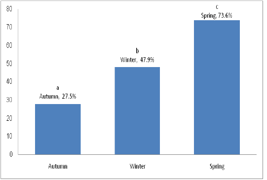
Figure 1: Infection rate according to year-season in sheep affected with gastro-intestinal parasites in Kirkuk, Iraq. Different letters (i.e., a, b, c) means significant differences at P< 0.05.

Figure 2: Infection rate according to age-groups in sheep affected with gastro-intestinal parasites in Kirkuk, Iraq. The star (*) means significant differences at P< 0.05.
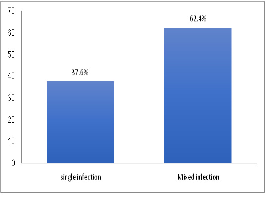
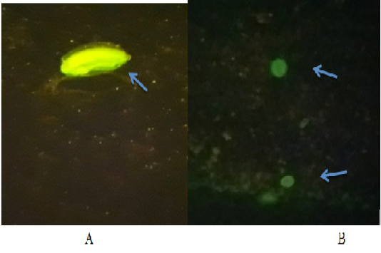
Figure 4: Fluorescing gasto-intestinal parasites egg using acridine orange stain, examined under fluorescent microscope 20X, A: Nematodes eggs; B: Eimeria spp.oocyst
Discussion
In current study, 85 samples were positive for gastrointestinal parasites (i.e., infection rate 53.1%). This rate is close to that documented in Mosul (54%) by Al-Bayati and Arsalan (2009), and lower than that recorded in Baghdad (75.1%) by Fadl et al. (2011), and Diyala (86.7%) by Minnat (2014). These differences could be attributed to the variation in the environmental and geographical conditions, time of sample collection, sampling method and technique.
The highest infection rate of gastrointestinal parasites was recorded in Spring (73.6%). High parasitic infection rate during Spring could be attributed to warm and humid weather, in addition to high rainfall, which provides suitable environment for high infection (Fadl et al., 2011; Hasan and Abed, 2012).
In current study, sheep with age-group 1-2 years-old showed higher infection rate compared to other age-groups. Age is considered an important risk factor for gastrointestinal helminthiasis (Raza et al., 2007). The low infection rate in adult sheep could be attributed to repeated exposure to the parasites leading to acquired immunity against parasitic infections and expelling the parasite from the body (Tesfaheyw et al., 2013; Dagnachew et al., 2011).
The infection included both single (37.6%) and mixed (62.4%) infection with more than one type of parasites. The high percentage of mixed infection could be associated with contamination of pastures with the eggs of different parasites, and the susceptibility to infection due to immune status of infected animals.
In this study, numerous parasites eggs were identified. Eggs of Marshallagia marshalli were dominant among eggs of the other species. This is consistent with that documented in (north of Iraq) Mosuland Garmiyan by (Al-Bayati and Arsalan, 2009, Al-Zandee et al., 2016), but differ from that of Fadl et al. (2011) and Kadhim, (1972) who recorded Haemonchus contortus as the most prevalent species in (Middle of Iraq) Baghdad. The differences in the infection ratios between the current study and other due to the variation in the number of samples and geographical differences among Southern, Middle and Northern regions of Iraq.
The study indicated the presence oocysts of Buxtonella sulcata in the examined sheep’s feces. This is consistent with (Solusby, 1986; Sulaiman et al., 2005). B. sulcata is naturally found in the intestines of rats and pigs, and transmitted to human and other animals through contamination of the food by the parasites oocyst causing inflammation of the intestine and watery diarrhea (Sulaiman et al., 2005).
In this study, eight species of Eimeria oocysts (27%) were diagnosed in the faeces of infected sheep. This result is different from that documented in Iraqi provinces, it was lower than the rate recorded in Baghdad: (49%) (Fikru et al., 2006), Diyala: (86.09%) (Minnat, 2014), Mosul: (60.5 to 63.6%) (Sulaiman et al., 2005; Ali et al., 2005), Sulaimanyah: (72%) (Kareem and Yücel, 2015) and Garmiyan (31.3%) (Al-Zandee et al., 2016). On the other hand, the infection rate was higher than that recorded in Erbil 3.25% by Ahmed et al. (2015) and in Kirkuk 18 to 36.2% by (Ali et al., 2005; Hassan and Barzinji, 2018). This difference could be due to the variation in the period of sample collection and diagnostic methods between our study and others.
The infected sheep exhibited numbers of clinical signs. The harmful effect of parasites can cause changes in the mucosal lining of gastrointestinal tract,which can lead to decrease absorption of nutrients and increase secretion of fluids causing signs like anorexia, weakness, pale mucous membrane, easily detach of wool, diarrhea and dehydration (Radostits et al., 2007). Loss of wool can be a complication of malnutrition which effect in the growth of wool follicles (Radostits et al., 2007).
Acknowledgments
This study was supported by the Department of Internal and Preventive Medicine, College of Veterinary Medicine, University of Mosul, Mosul, Iraq.
Conflict of Interest
The authors declare no conflicts of interest.
Authors Contribution
All authors contributed equally.
References


