Advances in Animal and Veterinary Sciences
Research Article

Clinical Dystocia in Iraqi Buffaloes in Mosul City
Mohammed A. Rahawy*
Department of Surgery and Theriogenology, College of Veterinary Medicine, University of Mosul, Iraq.
Abstract | In this study, dystocia weather maternal and / or fetal causes in Iraqi cow-buffaloes were analyzed. A total of 35 cow-buffaloes were admitted to the Clinic College of Veterinary Medicine, Mosul University 12 to 24 hours after starting delivery. The cow buffaloes suffering from dystocia. Either maternal or fetal (37.1%) in origin (62.85%), The maternal one showed a prevalence of 22.85%, 20%, 14.28%, 5.72% due to primary uterine inertia, incomplete dilation of a cervix, uterine torsion and narrow pelvis in buffaloes respectively. While the fetal causes showed a prevalence of 22.85%, 20%, 14.28%, 5.72% due to primary uterine inertia, incomplete dilation of a cervix of 17.15% due to maldisposition in cow- buffaloes as compared to fetal monster and fetal emphysema. 12.8%, 8.58% respectively.
Keywords | Buffaloes, Clinical Dystocia, Uterine Torsion, Incomplete cervical dilation, Fetal maldisposition.
Received | February 04, 2019; Accepted | April 24, 2019; Published | July 03, 2019
*Correspondence | Mohammed A. Rahawy, Department of Surgery and Theriogenology, College of Veterinary Medicine, University of Mosul, Iraq; Email: mohammedrahawy@yahoo.com
Citation | Rahawy MA (2019). Clinical dystocia in iraqi buffaloes in mosul city. Adv. Anim. Vet. Sci. 7(8): 715-719.
DOI | http://dx.doi.org/10.17582/journal.aavs/2019/7.8.715.719
ISSN (Online) | 2307-8316; ISSN (Print) | 2309-3331
Copyright © 2019 Rahawy. This is an open access article distributed under the Creative Commons Attribution License, which permits unrestricted use, distribution, and reproduction in any medium, provided the original work is properly cited.
Introduction
Failure to progress in labor, either maternal and/or fetal causes, for instance, an obstruction or constriction of the birth passage or abnormal size, shape, position, or condition of the fetus, or pathological or difficult labor is referred to dystocia (Lombard et al., 2007; Zaborski Zaborskiet al., 2010; Uzamy et al., 2010). Despite of 1-2% of incidence rate of dystocia in river buffalo. But still impact negatively on buffalo production and reproduction (Jainudeen,1986; Kaushik et al., 2005; Mee et al., 2011). Case reports of field cases and/ or cases admitted to veterinary clinics are the essential database recorded data for calculating the incidence rate of dystocia (Phogat et al., 1992; Purohit and Mehta, 2006; Ali, 2008). Factors affecting on dystocia can be summarized into two main categories: genetic and non-genetic factors, which intern classified as direct factors and phenotypic factors related to calf and cow (Mee, 2008). Mal presentations, uterine torsion, body weight at birth, twines, perinatal death, ring womb, birth body condition at calving and duration of gestation period are referred to direct and phenotypic factors. In addition to that, age, season of calving, calf gender, nutrition, hormonal status of cow-buffalo at pre parturient time also being consider as factors affecting on dystocia as well as the genetic factors (Mee, 2008; Uzamy et al., 2010). Fetopelvic discrepancy is the top most causes of dystocia in buffalo heifers compare with repeatedly cases of fetal disposition’s cases in pleuriparous cows (Bellows et al.,1996). For instance, maternal pelvic size and calve body mass at delivery times are classified to fetopelvic disproportions (Berry et al., 2007). A higher percentage of maternal dystocia was recorded in buffalo rather than cow as well as fetal dystocia had been reported by (Arthur et al., 1996; Nanda et al., 2003; Ettema and Santos, 2004). Complications during delivery in a birth canal almost produce uterine torsion except congenital anomalies in a birth canal (Ettema and Santos, 2004). The fetal monstrosity’s rates are higher degree than hereditary deformities that been recorded in river buffalo and this rate still within minor rate due to lack in registered cases in veterinary clinics (Sharma et al., 1996). 7.4% of neonate anomalies were classified as fetal causes of dystocia cases in Brazilian cow-buffalo, especially in the Murrah buffalo’s breeds and classify as congenital anomalies (arthrogryposis, myotonic, and mechano-bullous genodermatoses) (Singla, and Sharma,1992). Different etiologies such as metabolic disorders, anatomical and functional anomalies can be classified as hereditary anomalies of a birth canal in buffalo (Purohit, 2006). Insufficient cervical dilation is rare in cow-buffalo as a cause of maternal dystocia due to the delivery position of an animal and it’s less than 5.1% of total cervical dystocia reasons in buffalo compare to high rate of ring womb in cattle (Berger et al.,1992; Arthur et al.,1996; Marcolongo et al., 2010).
Right sided with uterine torsion is more frequently than left sided torsion, which is considered as normal records due to a high percentage of pregnancy in a right side of uterine horn in buffalos, partially packed rumen plays an important role in left sided torsion but in rare cases of uterine torsion (Wehrend, and Bostedt, 2003; Das et al., 2008). Although twin pregnant buffalo is rare, also in such a case, the urine torsion is rare too (Purohit and Mehta, 2006) but in cattle, these rare cases are a lessor than in buffalo (Penny, 1999) with maximum occurrence during second to third calving (Siddiquee and Mehta, 1992). Inadequacy in strength of uterine contraction with normal cervical dilation leads to uterine inertia, which is a most primary causative factor of dystocia in buffalos (Matharu and Prabhakar, 2001), which in turn mainly due to hypocalcemia (Nanda, 1995). Other causes for uterine inertia may be due to muscle fatigue of a uterine musculature as a result of continues strong contractions subsequent to failure of delivery of a mal disposed or oversized fetus or due to obstruction in the birth canal (Pargaonkar et al.,1993).
In this study, both maternal and fetal cause of cow buffalo dystocia was evaluated in admitted cases to a veterinary clinic of Veterinary Medicine College of Mosul University in Mosul City in Iraq.
Material and Methods
The current study was carried out on a total of 35 cases suffering from dystocia in River Iraqicow-buffaloes presented to the Teaching Veterinary Clinical Service in Collage of Veterinary Medicine -Mosul University, and presented to Clinics of veterinary obstetrics in Mosul City, during the period of 2012 – 2018. The causes of Dystocia have been classified as maternal, fetal. Treatment procedures included allowing the determinate amount of time for the cow buffaloes to calve by it, therapy to raise myometrial tone and cervical dilatation, which includes supplementation of calcium, fluids and oxytocin hormone.
Results
All the cow buffaloes were accessible to the Veterinary Clinical 12 to 24 hours after the onset of fetal expulsion (second stage of labor). The incidence of different causes of dystocia is presented in Table 1. The incidence of dystocia due to maternal greater than fetal causes was (62.85% and 37.15% percent), the incidences of maternal cause of dys
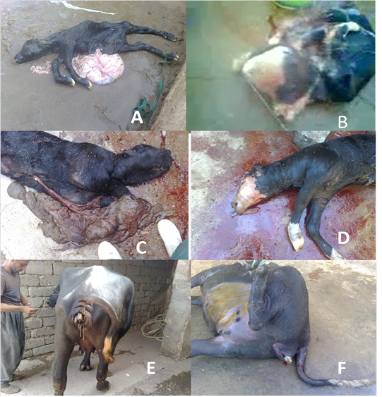
Figure 1: (A) Dead male buffaloes fetal Ascites after delivery by penetrated of abdominal wall and then lubrication and forced extraction the Fetus. (B) Dead buffaloes fetal Hydrocephalus with perosomuselumbis after delivery by Cesarean Section. (C) Dead male buffaloes fetal Anasarca after delivery by Cesarean Section.(D) dead fetal emphysema after delivery by Cesarean Section.(E) Cow-Buffalo suffering from Dystocia due to lateral deviation of the head delivered after correction of the head.(F) Cow- Buffalo suffering from Dystocia due to Incomplete cervical dilatation delivered by Cesarean Section.
Table 1: Percent Incidence of different type of dystocia in Iraqi cow-buffaloes
| Maternal causes | Number of case | Percentage % |
| Primary Uterine inertia | 8 | 22.85% |
| Incomplete cervical dilation | 7 | 20% |
| Uterine torsion | 5 | 14.28% |
| Narrow pelvis | 2 | 5.72% |
| Total | 22 | 62.85% |
| Fetal causes | Number of case | Percentage % |
| Fetal mal disposition | 6 | 17.15% |
| Fetal monster | 4 | 11.42% |
| Fetal emphysema | 3 | 8.58% |
| Total | 13 |
37,15 |
tocia were Primary Uterine inertia (22.85) but incomplete dilation of cervix (20%), uterine torsion (14.28) and Narrow pelvis in buffaloes (5.72%). While the main fetal cause was mal disposition of the fetus in Iraqi cow buffaloes (17.15%),because of lateral deviation of the head, downward displacement of the head, bilateral carpal flexion, bilateral shoulder flexion, bilateral hock flexion. While teral hip flexion but the fetal monsters due to conjoined twins were attached tetrabrachius), cephalus dipustetrabrachis), Hydrocephalus with perosomuselumbis, anasarca and fetal ascites in Iraqi cow -buffaloes, While the incidence of fetal emphysema was 8.58% in cow-buffaloes (Figure 1).
Discussion
Most of the recorded data in India showed that 4.6% - 5.4% in Surti, 5.6%-12.6% in Murrah and 8.94% in Jaffarabadi buffalo breeds were suffered from dystocia regardless of the etiology of the case (Biggs and Osborne, 2003). Low incidence rate of dystocia cases in buffalo compared to dystocia rate in cattle could be due to variation in anatomic aspect of a birth canal in these animals as well as birth body mass of fetus and calving positions (Kodagali, 2003). Such anatomic variations in a birth canal could be the main reason for having 20-70 minutes calving periods as first – second stage of calving in buffalo, which is recorded by (Pargaonkar et al., 1993; Agarwal and Tomer, 2003; Khan et al., 2009). Jainudeen, (1986) he refers to dystocia in buffalo was not big issue in water buffalo, despite the incidence rate of dystocia in swamp buffalo lesser than river buffalo and Pleuripara than Primipara breeds (Mody et al., 2002).
The study results showed that maternal and fetal causes as 62.85%, 37.15% respectively in admitted cases was in agreement with previous work by (Majeed, 2001; Ramasamy and Singh, 2002). Recorded maternal causes in the current study such as primary uterine inertia (22.85%) due to twin pregnancy, pre-calving milk fever and high body mass of newborns is the main reasons for uterine inertia. Furthermore, cervical dilation happens with normal positioning and posture of a fetus presented during delivery but lack of uterine contraction due to various reasons leads to dystocia problems. 59.16% and 40.84% rate of dystocia incidence in Murrah buffalo because of maternal causes was in agreement with current results (Matharu and Prabhakar, 2001). Major frequent reason for primary uterine inertia in buffalo is the progressions of birth without continue to the second stage of labor (Siddiquee and Mehta, 1992). (Phogat et al.,1992) Disagree with our recorded data of cases showing hypocalcemia and exhibited signs of milk fever at the beginning of calving, which related to 5.9% of uterine inertia incidence rate. Secondary uterine inertia happens as results of exhaustion consequent on prolonged dystocia (Berger et al., 1992; Srinivas et al., 2007). Fatigued uterine musculature due to failure to delivery or heavy muscle contraction with abnormal delivery either maternal and / or fetal cause is another factor leads to secondary uterine inertia accompanied with oversized fetus, birth canal obstruction. Depletion of calcium due to continuous uterine contractions without compensation from the body as a result of the low level of calcium or lack of time, to compensate despite the good level within the body will lead to weak uterus and failure to push out the fetus out of the birth canal normally. In hypocalcemic buffalo, delivery needs expulsive forced contraction to help the uterine contraction to push the fetus through the birth canal, while the animal is hypocalcemic, the strength of muscle contraction is insufficient to have a role in second stage of labor to expel the fetus (Biggs and Osborne, 2003). Failure of abdominal wall muscles to contract forcefully to contribute in the second stage of labor might be visceral pain, urinary tract infection, weak and / or tired abdominal muscles, which could be a presence in old age animals as well as traumatic reticulitis/pericarditis, painful conditions of the diaphragm (Arthur et al., 1996). 20% of admitted dystocia cases showed partial cervix dilation according to the clinical examination via vaginal inspection; this result is combatable with (Ramasamy and Singh, 2002) who found that 13.10% of dystocia cases in Murrah buffalos were suffered from insufficient cervix dilation.
Failure of a cervix to dilate completely might be due to mechanical, functional and /or hormonal cause’s hormonal cause. Cervical dilation undergoes properly mostly by hormonal control and enzymatic reactions in the smooth and straight muscles of a cervix as well as birth canal, which control by estradiol plus prostaglandin F2α (Sharma et al., 1992; Jackson, 1995). In addition to the mechanical forces of uterine muscle contractions and passes of fetal through the uterus outward to a cervix play an important role in sufficient cervix dilatation throughout labor in the buffalo, activation of a non-pathological inflammatory process has another role in opening cervix normally, which is still unknown mechanisms could be its inhibition leads to dystocia (Rajabi et al., 1988).
In the current study, a 14.28% of studied cases found to be suffered from uterine torsion, which is another cause of buffalo’s dystocia and has a higher ratio than cattle. Uterine torsion is the highest ratio of dystocia causes among the admitted cases, which is in agreement with (Purohit et al., 2011) who, reported that buffalo case with uterine torsion was more advanced than those in cattle and most frequent in river than swamp buffalos. Pleuriparous cow’s buffalo defined as the greater incidence rate of uterine torsion with major ratio of dystocia during second and third calving with right sided torsion (Nanda et al., 1991; Singh, 1995; Matharu and Prabhakar, 2001). Left side uterine torsion might be happen when the rumen was partially filled and the degree of torsion in most cases a round 90° -180° which almost lead to rapid fetal death and uterine adhesions with visceral organs could be developed, uterine torsion must be considered emergency cases in such cases (Prabhakar et al.,1994).
Another cause of dystocia is pelvic defects weather is congenital and / or due to traumatic defects, plays an important role in labor. In the current study 5.72% of studied cases of Primiparous buffaloes had narrow pelvis signs that lead to dystocia in admitted cases. Hereditary causes of pelvic defects, for instance, small size and incomplete pelvic ligament’s development or small-sized breeds with outsized fetuses could be other reasons for dystocia due to narrow or defected pelvis. Outsized fetus can results in obstruction of a birth canal due to small-sized pelvis or abnormal pelvis of the cow-buffalo (Purohit and Mehta, 2006). Sacral luxation or displacement was recorded as a small bony pelvis cause added to genetic causes of a small-size pelvis (Biggs and Osborne, 2003). Bovine Primipara found to have a higher rate of an asymmetrical pelvis size around 7.79% of registered cases with a narrow pelvis dystocia cause (Purohit, 2006). 9.2% of buffalo submitted with dystocia had a narrow pelvis which recorded by (Arthur et al., 1996).
Lateral deviation of the head is the frequent fetal mal disposition in dystocia cases in buffalo (17.15%) as well as, head displacement, bilateral carpal and / or shoulder flexion, hip and hook bilateral flexion too. Most of these fetal mal positions are probably due to failure of a fetus to rotate normally inside the uterus in addition to reduced viability of the offspring. Failure of the fetus to rotate from the intrauterine position to the normal parturient position may result in dystocia (Phogat et al., 1992). Anomalous fetal presentations at time of calving add 1%-5% of total dystocia cases (Purohit et al., 2012).
Mild developments or abnormalities in embryogenesis results in anatomical deformities in the fetuses referred as monstrosities, which play another role in dystocia causes and are common in the buffalo. Monstrosity is a developmental disorder that associates with various organs and systems, which can cause great distortion of the individual. Maternal and genetic factors play an important role in fetal anomalies in early stage of cell differentiation and / or during embryogenesis, and these anomalies develop later into monster fetuses leading to dystocia in most cases. 7.9% - 12.8% of admitted cow-buffalo suffered from monstrosities and major percentage of monstrosities were recorded in river buffalo rather than swamp buffalo (Garrousi, 2004). Congenital deformities like conjoined twins were fused in their thoracic regions (thoracopagus), had four front legs (tetrabrachius), four hind legs (tetrapus) and two separate tails (dicaudatus) with single head and neck referred as a monocephalus thoracophagus tetrabrachius tetrapusdi caudatus twin monster. Other deformities arehydrocephalus, anasarca and foetal ascites, the incidence rate of thesemonstrosities 12.8% and 8.58% with fetal emphysema, and current finding agree with study results found by (Purohit et al., 2012).
Conclusion
Dystocia cases are stressful events for both mother and offspring with potentially lifelong consequences and have a large economic impact on farmers due to calf death, injury or death to the cow buffaloes, veterinary cost, as well as the decrease pregnancy rate of the dam after losing a calf. The incidence of dystocia due to maternal with primary uterine inertia and incomplete dilation of the cervix higher than mal disposition appears to be more frequent of fetal causes in Iraqi buffaloes. It is commonly in first-calf heifers of buffaloes.
Acknowledgements
The project was approved by the scientific committee of Mosul University for animal research and animal welfare, and was fully supported by the Faculty of Veterinary Medicine of University of Mosul. The authors are indebted to the buffaloes owners for their endlees colaboration.
References


