Advances in Animal and Veterinary Sciences
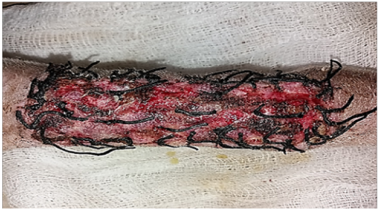
Gross image of autograft site of dog’s leg after one week of surgery. Showed cardinal signs of inflammatory reaction included swelling and redness (due to hemorrhage) with an area of necrosis and sloughing of the dermis layer.

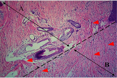
Histological section of autograft site of dog’s leg after one week of surgery. Showed two sides of the tissue, the original tissue showed no signs of rejection (A), while the grafted tissue (B) showed necrosis and degeneration of the dermis layer (arrowhead). H&E, 100x.

Gross image of autograft site of dog’s leg after one month of surgery. Showed formation of granulation tissue (scar) with the restoration of normal skin color without hair formation.
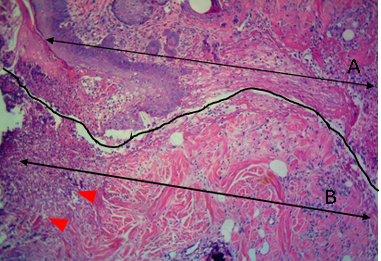
Histological section of autograft site of dog’s leg after one month of surgery. Showed original tissue (A) and grafted tissue (B). Extension of collagen fiber to connect the wound sides (arrowhead). H&E, 100x.
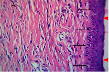
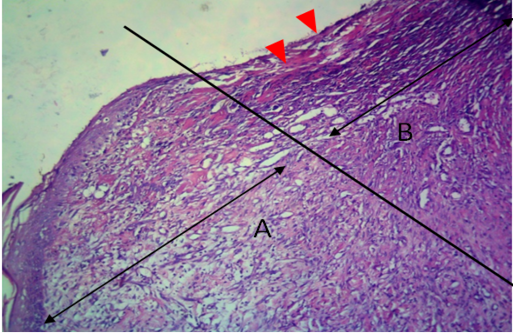
Histological section of autograft site of dog’s leg after one month of surgery. Showed grafted tissue (A) with the restoration of the epidermis layer (arrowhead) and formation of spongiosum layer (arrow).
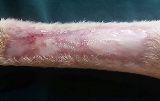
Gross image of autograft site of dog’s leg after two months of surgery. Showed formation of granulation tissue (scar) with the restoration of normal skin color without hair formation.
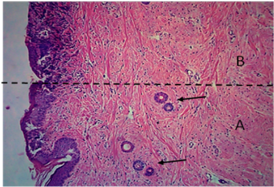
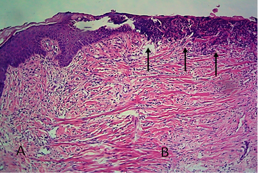
Histological section of autograft site of dog’s leg after two months of surgery. Showed original tissue (A) and grafted tissue (B), the main differences between the two sides of tissue was the presence of hair follicle and sweat glands (arrow). H&E, 100x.

Gross image of allograft site of dog’s leg after a week of surgery. Showed inflammatory signs, areas of hemorrhage, necrosis and skin ulcers.
Histological section of allograft site of dog’s leg after a week of surgery. Showed original tissue (A) and grafted tissue (B). Necrosis and sloughing of the epidermis layer of grafted tissue (arrowhead). H&E, 100x.
Gross image of allograft site of dog’s leg after a month of surgery. Showed granulation tissue formation with the absent of gross signs of tissue rejection.
Histological section of allograft site of dog’s leg after one month of surgery. Showed original tissue (A) and grafted tissue (B). Necrosis and hemorrhage present in epidermis layer of grafted tissue (arrowhead). H&E, 100x.

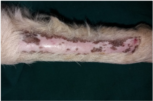
Two-months post grafting showed absent signs of rejection little hair formation and pigmentation like the donor skin
Histological section of allograft site of dog’s leg after two months of surgery. Showed original tissue (A) and grafted tissue (B), also granulation tissue formation in grafted tissue.


