Advances in Animal and Veterinary Sciences
Case Report

A Veterinary Clinical Case of Severe Chronic Haemonchus contortus Infection in a Goat: The Clinical Management of the Case and Pathology Findings
Faez Firdaus Abdullah Jesse1, 2, 3*, Eric Lim Teik Chung1, 4, Yusuf Abba5, Mohd Azmi Mohd Lila2, Siti Nor Aishah2, 3, Syahirah Affandi2,3, Asinamai Athliamai Bitrus6, Innocent Damudu Peter2, 5, Idris Umar Hambali5
1Institute of Tropical Agriculture and Food Security, Universiti Putra Malaysia, 43400 Serdang, Selangor, Malaysia; 2Department of Veterinary Clinical Studies, Faculty of Veterinary Medicine, Universiti Putra Malaysia, 43400 Serdang, Selangor, Malaysia; 3University Veterinary Hospital (UVH), Faculty of Veterinary Medicine, Universiti Putra Malaysia, 43400 Serdang, Selangor, Malaysia; 4Department of Animal Science, Faculty of Agriculture, Universiti Putra Malaysia, 43400 Serdang, Selangor, Malaysia; 5Faculty of Veterinary Medicine, University of Maiduguri, P.M.B 1069 Maiduguri, Borno Nigeria; 6Research Unit, Microbial Food Safety and Antimicrobial resistance, Department of Veterinary Public Health, Faculty of Veterinary Science, Chulalongkorn University, 10330 Pathumwan Bangkok, Thailand.
Abstract | This case report describes the clinical management and pathology findings of a case of severe chronic Haemonchus contortus infection in a goat. An adult male Jamnapari goat weighing 40kg was presented with the primary complaint of depression and recurrent diarrhea. Physical examination findings revealed the buck was emaciated with a poor body score of 1 out of 5, dull and depressed, severe pale mucous membrane with the FAMACHA score of 5, and greenish faecal stain at the perineal region of the buck. Diagnostic and laboratory findings revealed that the buck had 3400 epg count which was significant value for Haemonchus contortus infection, PCV of 9%, blood analyses revealed anaemia and hypoalbuminemia. The case was managed medically with instituting blood transfusion, anthelmintic, antidiarrheal, iron supplement,and NSAIDs. Post-treatment progress showed the condition of the buck was not improving. The buck was euthanized and post mortem was conducted. Gross post mortem findings of this case were thickening of the wall of abomasum indicates due to severe and chronic infection by Haemonchus contortus, presence of frothy exudates at thoracic inlet, airways and lung parenchyma indicates pulmonary oedema due to hypoalbuminemia, and hepatomegaly together with loss of subcutaneous and visceral organs fat were observed in this case may due to the chronic malnutrition and Haemonchus contortus infection. Therefore the cause of death of this buck, in this case, was due to respiratory and circulatory system failure because of pulmonary oedema and hypoxia as a result of hypoalbuminemia and anaemia caused by severe Haemonchus contortus infection and malnutrition.
Keywords | Veterinary, Haemonchus contortus, Clinical Management, Pathology Findings, Goat.
Received | February 28, 2019; Accepted | March 30, 2019; Published | April 23, 2019
*Correspondence | Faez Firdaus Abdullah Jesse, Institute of Tropical Agriculture and Food Security, Universiti Putra Malaysia, 43400 Serdang, Selangor, Malaysia; Email: jesse@upm.edu.my
Citation | Jesse FFA, Chung ELT, Abba Y, Lila MAM, Aishah SN, Affandi S, Bitrus AA, Peter ID, Hambali IU (2019). A veterinary clinical case of severe chronic haemonchus contortus infection in a goat: the clinical management of the case and pathology findings. Adv. Anim. Vet. Sci. 7(6): 503-507.
DOI | http://dx.doi.org/10.17582/journal.aavs/2019/7.6.503.507
ISSN (Online) | 2307-8316; ISSN (Print) | 2309-3331
Copyright © 2019 Jesse et al. This is an open access article distributed under the Creative Commons Attribution License, which permits unrestricted use, distribution, and reproduction in any medium, provided the original work is properly cited.
Introduction
Helminths infection is rampant in livestock farming especially small ruminants due to grazing activities on pasture contaminated with third stage infective larvae of parasitic nematodes (Chandrawathani et al., 2009). In Malaysia, the main species causing helminths infection are Haemonchus spp., Trichostrongylus spp., and Oesophagostomum spp (Jesse et al., 2017a,b). According to Nor-Azlina et al. (2013), Haemonchus contortus (H. contortus) which also known as ‘barber pole’ worm is the most pathogenic nematode that feeds on blood. H. contortus is one of the most fecund strongyle nematodes where individual females are capable of producing thousands of eggs per day that can lead to rapid larval pasture contamination and associated outbreaks of haemonchosis. In small ruminants, Haemonchosis is the second most important cause of mortalities in small ruminants in Malaysia (Nor-Azlina et al., 2013).
H. contortus infection can be manifested as acute or chronic disease where the main clinical signs observed during acute condition are haemorrhagic anaemia, dark-coloured faeces, oedema, weakness, reduced muscle mass or sometimes sudden death occur where else in chronic condition decreased food intake, weight loss and anaemia are the most common clinical signs were observed (Jesse et al., 2015; Abdullah et al., 2016). Basic principles in the therapeutic management of managing clinical cases of haemonchosis are to correct the anaemia and dehydration condition, managing diarrhea and weakness (Jesse et al., 2015). The most common gross lesions observed in the cases of haemonchosis of small ruminants are generalized oedema, anaemia, and presence of fluid in the body cavities. For abomasal contents, there will be fluidal and sometimes mixed with free blood in the cases of a large number of adult H.contortus parasites. Lesions such as ulcerative haemorrhagic spots can be observed on the abomasal mucosa with hyperaemia at the abomasal folds where the parasites found to adhere. Microscopical examination of the abomasum will reveal extensive haemorrhages at the mucosa and submucosa with infiltration of eosinophils and mononuclear cells and hyperactivity of the goblet cells (Iqbal et al., 1993; Pathak et al., 2014; Dutta et al., 2017).
For prevention, the appropriate strategies can be implemented that includes animal management programmes to avoid excessive H.contortus challenge, genetic and nutritional approaches that will enhance the resistance and resilience to infection, and monitoring program towards H.contortus infection for individual animal or herd basis. For anthelmintic resistance management, the appropriate use of effective anthelmintics and refugia-based treatment schedules should be implemented. Vaccination against H. contortus can opt as another option that appears to have significant potential in control against H. contortus infection (Besier et al., 2016). In this veterinary clinical case report describes the clinical management and pathology findings of a veterinary clinical case of severe chronic H. contortus infection in a goat.
Veterinary Clinical Case Report
History: An adult male Jamnapari goat weighing 40kg was presented to University Veterinary Hospital (UVH) UPM with the primary complaint of depression and recurrent diarrhea. From the history obtained the buck had a history of H. contortus infection past two years with anthelmintic resistance and the buck was managed semi-intensively and graze at the same pasture plot.
Physical Examination and Diagnostic Work: Physical examination findings revealed the buck was emaciated with poor body score of 1 out of 5, dull and depressed, severe pale mucous membrane with the FAMACHA score of 5 and greenish faecal stain at the perineal region of the buck. Diagnostic and laboratory findings revealed that the buck had 3400 epg count which was significant value for H.contortus infection, PCV of 9% indicate the buck was having severe anaemia that explains the severe score of FAMACHA, for Complete Blood Count (CBC), the results showed anaemia of normocytic hypochromic with leukocytosis characterized by monocytosis and neutrophilia with a left shift. The blood biochemistry results revealed hypoproteinemia, hyperkalemia, uremia, hypercreatinaemia, hypoalbuminemia, increased in creatinine kinase and albumin globulin ratio. From the history and physical examination findings supported with diagnostic aid, the buck was diagnosed suffering from severe chronic H.contortus infection.
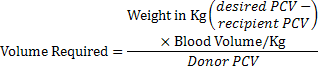
Clinical Management: The case was managed medically with instituting a blood transfusion to improved PCV from 9% to 15% and total of 436ml blood was transfused on the first day of hospitalization using formula stated by Kahn and Line (2005), where 436 mL of blood was collected from a donor goat with PCV 33% (formula as below). For the first 15 minutes, the blood was transfused at a rate of 1drop/second to monitor for adverse reaction and it was increased to 2 drops/second until all the required amount of blood was transfused.
For other therapeutics management of the case maintenance fluid of NaCl (0.9%) was instituted for first 2 days of hospitalization, anthelmintic Levamisole (2.5 ml/kg) PO once was given as dewormer of choice, antidiarrheal kaolin-pectin (30ml/25kg) PO TID for 5 days, iron supplement Fercobsang (5ml/adult goat) intramuscular injection once for 5 days and pain management using NSAIDs of Meloxicam (0.2 mg/kg) subcutaneous injection once for 5 days were planned as the therapeutic regime for this buck. Post-treatment progress showed the buck was not improving where the PCV remained consistently at 13% post blood transfusion for 5 days without any improvement, the worm count showed an increased in the worm count where 3950 epg was recorded on the fifth-day post-treatment, the buck was weak and on sternal recumbency. Additional diagnostic aid was planned for this buck where the chest radiography was done and the result revealed that the buck had a presence of fluid in the thoracic region. From the clinical judgement, the buck was having a poor prognosis and to ease the pain and welfare of the buck clinical decision of euthanasia were opted in this case. The buck was euthanized and post mortem was conducted.
Postmortem Findings: Gross post mortem findings of this case were the carcass was observed to have poor body condition with minimal visceral and subcutaneous fat with a pale appearance of serous membrane. Approximately 400mL of blood tinged fluid in the thorax (hydrothorax) region was collected from the post mortem examination. Presence of frothy exudate at the thoracic inlet, airways, and parenchyma of the lungs and thickening of the wall of abomasum were also observed. Generalized severe serous atrophy of fat was observed in the omentum, kidney, and heart. The pericardium and the kidney capsules were both pale. The liver was mildly enlarged and firm (Figure 1).
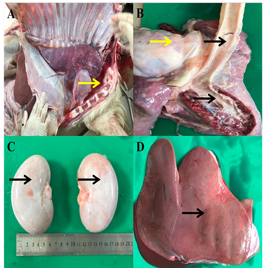
Figure 1: (A) Presence of blood tinged fluid in the thoracic cavity (B) Presence of frothy exudates (black arrow) Thickened pericardial sac (yellow arrow) and (C) Pale kidney capsules (D) Mild enlargement of liver and firm with rib imprints.
Therefore to summarize the gross post mortem findings of this case are thickening of the wall of abomasum indicates due to severe and chronic infection by H. contortus, presence of frothy exudates at thoracic inlet, airways and lung parenchyma indicates pulmonary oedema due to hypoalbuminemia and hepatomegaly together with loss of subcutaneous and visceral organs fat were observed in this case may due to chronic malnutrition and H.contortus infection. Therefore the tentative cause of death of this buck, in this case, was due to respiratory and circulatory system failure due to pulmonary oedema and hypoxia due to hypoalbuminemia and anaemia due to severe H.contortus infection and malnutrition.
Discussion
Haemonchosis is a disease of small ruminants that will results in severe anaemia and death if prompt intervention is not done in early stages of the disease (Jesse et al., 2015; Mohammed et al., 2016). In this current case report, the buck had a history of 2 years recurrent chronic haemonchosis infection. This may be associated with either poor environment that has been contaminated with high infective larvae or humid tropical environment in Malaysia which favor year-round development and survival of pre-parasitic stages of H.contortus on pastures (Dorny et al., 1995).The history from this case in agreement with Dorny et al. (1995), where the buck was managed semi-intensively and graze at the same pasture plot which gives higher chances and percentage of reinfection by H.contortus.
Anthelmintic resistances to all the major anthelmintic drug classes have been reported throughout the world in small ruminant production in countries such as South America, South Africa, Malaysia and the USA (Kaplan, 2004). Chandrawathani et al. (1999), stated that 34% of the small ruminant farms from the northern state of Peninsular Malaysia were resistance towards benzimidazole and in the year of 2004, the same author reported totalantihelmintic failure in Sabah, Malaysia. Chandrawathani et al. (2004), mentioned that anthelmintic resistance can occur due to the unnecessary practice of drenching and goats metabolize anthelmintics faster than sheep, thus the same dosage is less efficacious hence promoting resistance to nematodes in goats. In this case report, the animal was a goat and this can be a contributing factor towards the anthelmintic resistance developed in this buck that leads to treatment failure.
Proper nutrition can increase the ability of the host’s resistance by limiting the establishment, growth rate and parasite population and where else poor nutrition reduces the ability of the host immune system to counter the effects of parasitism (Mohammed et al., 2016). The buck, in this case, had severe malnutrition and body score that maybe attributed and aggravates toward the H.contortus infection. The prognosis of the buck was poor may due to chronic diarrhea that may results in loss of vital nutrients that help in rejuvenation of the body and subsequent weight gain and lead to the leakage of plasma from the intestine that resulted in hypoproteinaemia and hypoalbuminaemia that were observed in this case (Abdullah et al., 2016).
FAMACHA chart is a visual estimation of the colour of the conjunctiva mucous membrane to assess the severity of anaemia (Malan et al., 2001; Glaji et al., 2013). The buck in this case report had a FAMACHA chart score of 5, which is a very poor score an indication of blood transfusion needs to be performed. However, the PCV remained at 13% unchanged throughout 5 days post-transfusion and this may due to normocytic hypochromic anaemia observed in this case. Zucker et al. (1974), stated that patients with hypochromic anaemia may have symptoms such as lack of energy and shortness of breath and thesesymptomswere in accord with the clinical signs observed in the buck in this case. Besides, the unchanged PCV value in this case post blood transfusion may due to hypo-regenerative anaemia where there is alteration of bone marrow progenitor cells and the cause may due extrinsic factor of parasitic infection stated by (De Cruchy et al., 1978; Jandl et al., 2003) as recorded in this case report. Another possible cause of non-responsive PCV value, in this case, may due to normocytic anaemia that was recorded in this case where the cause of normocytic anaemia in this buck due to nutritional anaemia that will have deficiency of vitamin B12, folic acid, and iron and this will aggravates the hypo-regenerative anaemia and that was observed in this case (Tefferi, 2003). The gross pathology findings in this case report are in accord with (Pathak et al., 2014; Dutta et al., 2017) where all the findings relate with severe H.contortus infection and aggravates with malnutrition condition of the buck in this case and this explains the poor prognosis of the case in this report.
H.contortus infection can be prevented and controlled in small ruminant farms by implementing short-term rotational grazing strategy or practicing full intensive management system and regular check of FEC and proper deworming protocol should be instituted in order to reduce the incidence of anthelmintic resistance (Chandrawathani, 2004). In the future application of Barbervax® vaccination practice may help in prevention strategy of H.contortus infection (De Matos et al., 2017).
Conclusion
In this veterinary clinical case report describes the clinical management and pathology findings of a veterinary clinical case of severe chronic H. contortus infection in a goat. The basic therapeutic and medical management, in this case, lead to no improvement due to the poor prognosis of the case as the buck was diagnosed having severe H. contortus infection aggravated with malnutrition condition of the affected animal. Therefore, prevention of this clinical case of severe chronic H. contortus infection from recurring in the affected farm can be done through proper feeding. The nutritional requirement needs to be reassessed and a holistic herd health program should be implemented.
Acknowledgements
The authors would like to thank the staff of the Department of Veterinary Clinical Studies, Universiti Putra Malaysia for their technical assistant.
Conflict of Interest
The authors declare that they have no conflict of interest.
Authors’ Contribution
All authors contributed equally and approved the final manuscript.
References


