Advances in Animal and Veterinary Sciences
Research Article

Therapeutic Effects of Hydro-Alcoholic Extract of Clove Buds and Econocapus Erictus on Infected Wounds Healing in Dogs
Ali I. Al-ameedi1*, Hussain Nahi2
1Department of physiology and pharmacology, College of Veterinary Medicine Al-Qasim Green University, Babylon- Iraq; 2Department of Surgery and Obstetrics, College of Veterinary Medicine Al-Qasim Green University, Babylon- Iraq.
Abstract | The aim of this study was to evaluate the effect of two plants hydro-alcoholic (ethanol) extracts on wounds infected with resistance Staph. Aureus as ointments and in vitro, by comparing their with 1% of penicillin ointment as control positive. Serial dilutions used to determine the MIC and MBC of extracts and penicillin.Thirty two mature street dogs were divided into four equal groups (control, treated one, treated two and treated three). In control group (C)applied penicillin ointments on the infected wound topically, treated one group (T1) received 16mg/ml of C. erictus as ointment during 3 days after wound infected.while treated two group (T2) the infected wound treated with 6mg/ml of clove buds extract as ointment. Treated three group (T3) same procedure mention above but treated with combination ointment (C. erictus ointment and clove ointment) in ratio 1:1. The results revealed that MIC and MBC of C.erictus, clove buds and penicillin were (8mg/ml-16mg/ml, 5mg/ml-10mg/ml and 2μg/ml-4 μg/ml) respectively. The clinical parameters included temperature, respiratory rate , and heart rates showed no significances (p≤ 0.05) between groups. The scar present large quantitate in control group when compared with other group, the lesser scar formation was visible in T3 group. Histopathological Examinations was done by taken the skin biopsies on 15rd, and 30th, day post wound infection, the histopathological results revealed that the wound healing in T3 group faster and better than in the other groups and the epithelial cells more maturity and the skin taken same normal shape in T3 group comparing with other groups. In conclusion we are demonstrate that clove buds and C. erictus hydro-alcoholic extracts have valuable pharmacological properties include antibacterial, anti-inflammatory and anti-pyritic in vitro and on infected wound with resistance Staph. aureus.
Keywords | Econocarpus, Clove, Ointment, Wound healing, Dog
Received | September 14, 2018; Accepted | November 01, 2018; Published | March 10, 2019
*Correspondence | Ali I Al-Ameedi, Department of physiology and pharmacology, College of Veterinary Medicine Al-Qasim Green University, Babylon- Iraq; Email: ali.alameedy89@yahoo.com
Citation | Al-Ameedi AI, Nahi H (2019). Therapeutic effects of hydro-alcoholic extract of clove buds and econocapus erictus on infected wounds healing in dogs. Adv. Anim. Vet. Sci. 7(5): 389-396.
DOI | http://dx.doi.org/10.17582/journal.aavs/2019/7.5.389.396
ISSN (Online) | 2307-8316; ISSN (Print) | 2309-3331
Copyright © 2019 Al-ameedi and Nahi. This is an open access article distributed under the Creative Commons Attribution License, which permits unrestricted use, distribution, and reproduction in any medium, provided the original work is properly cited.
Introduction
For years we have been looking for the ideal antibiotic, now it seems clear to us that the plants are so anti-containing of natural substances effective in the treatment of bacterial, fungal and viral infections. Medicinal plants are the wealthy bio-resource of drugs of conventional medicines, novel medicines, food supplements and classic medicines (Ncube et al., 2008; Nirmala et al., 2011). Although the synthetic chemistry still they have essential role in the discover and produce the drugs .the possibility of plant extracts have a good chance to yield new formulas for the prevention and cure many diseases (Kviecinski et al., 2008; Newman et al., 2003; Verpoorte, 2000) different active ingredient extracted from plants have been tested and recorded as new drugs patented like taxol, artemisinin and maprouneacin (Goodman and Walsh, 2001; Klayman, 1993; Carney et al., 1999) at the result of multi-targeting, safety and cheap of plant source products, there is a good chance to searching more and more for many plant agents and used as drugs. Martins et al. (2016) recorded many uses of plant published in folk medicine, it was reported to astringent, styptic and tonic preventing anemia, catarrh, conjunctivitis, diabetes, diarrhea, fever, gonorrhea, headache, hemorrhage, orchitis, prickly heat, swellings and syphilis. essential oils and their components are extensively used in medicine as ingredients in a diverse range of medical products, and in food manufacturing as flavoring additives. in addition to their antimicrobial properties, in food systems they can be considered as an extra intrinsic factor to increase the safety and shelf life of foods. they are also used in cosmetics as perfumes (Bakkali et al., 2008). Eugeniacaryophyllus (clove), belonging to the family myrtaceae, has several medicinal effects and its systemic and local use is widespread in traditional medicine. cloves are reported to have anti-oxidant (Shobana et al., 2000), anti-bacterial (Cai and Wu, 1996), anti-candida (Chami et al., 2004), local anesthetic (Ghelardini et al., 2001), anti-halitosis (Bakhtiarian et al., 2012), and aphrodisiac (Tajuddin et al., 2003) properties. Conocarpus erectus l., popularly known as man-grove button belongs to the family combretaceae and it is found in tropical and subtropical regions around the world (Bandeira, 2003). different parts as leaves, stem, fruits and flowers have antioxidant, anticancer and antimicrobial properties (Abdel-hameed et al., 2011). Staphylococcus aureus is one of the major human pathogens responsible for the infections of skin, soft tissue, respiratory tissue and bone joints, causing endovascular infections such as bacteremia, endocarditis, sepsis, and toxic shock syndrome (Krupanidhi and Vijaya 2017) The aims of this study was to investigate the antibacterial anti-inflammatory effect of two plants extraction in vitro and as ointments on infected wound with resistance Staph. aureus.
Materials and Methods
Preparation of the Extract
Conocarpus erectus: Fresh (Conocarpus erectus) leaves were collected from agricultural field in the College of Agriculture, Al-Qasim green university during September 2017. The leaves were washed under tap water 3 times, and then dried in room temperature at shade. The dried leaves were crushed by a laboratory blender.
Clove (Syzgiumaromaticum): 500 gram of the clove buds was taken from the market and make it powder by crushing with grinder.
Plant Extraction
Hydro alcoholic (ethanol) extraction of leaves of Conocarpus erectus and Clove buds powder was carried out according to (Harborne, 1984) by using 1000 ml flask in which 50 grams of leaves and buds powder was put in the flask, then up to1000 ml of 30% ethanol was added, mixed and extracted by magnetic stirrer at 40◦C for 48 hours Figure (1, 2 and 3), then filtered with gauze to get rid the residue then extra filtrated by whatman paper and millipore paper (0.5mm). The filterate dried up by using incubator at 40oC. The yield extract was calculated according to wet and powder plant leave extract yield according to the following equation and the final extract was kept frozen at –20◦Ċ until use.
Bacterial Isolate
1.5×10⁷ CFU/ml of Staph. aureus in physiological saline were obtained from the lab of microbiology of college of veterinary medicine at Al-Qasim green university. Table (1).
Table 1: Source of bacteria used in this study
| Bacteria | Characteristic | Source |
| Staphylococcus. aureus |
Oxacillin , cefoxitin⃰ |
Clinical isolate, College of Veterinary Medicine, Al- Qasim Green University |
*clinical isolate bacteria resistance to oxacillin and cefoxitin.
Minimum Inhibitory Concentration of Hydro-Alcoholic (Ethanol) Extracts against Staphaureus
MIC and MBC were performed as previously described (Halawani et al., 2011). Briefly extracts were serially diluted with nutrient broth to give series of concentrations in sterile microtitre plates. Each series of dilutions was inoculated with 1.5×10⁷ CFU/ml of Staph.aureus and incubated at 37o C for 18 hours before determining the least concentration that inhibited the appearance of visible growth. The minimum bactericidal concentrations (MBCs) were determined from broth microdilution assays by sub culturing 10 µl volumes from inhibitory concentrations onto Muller-Hinton agar plates.
Concentrations and Ointment Preparation
Depending on the results of MIC of plants extract and penicillin we choose the conc. Of each extract and penicillin, The plant extracts of clove buds and Conocarpus erectus leaves ointment and other constituent prepared by using (Vaseline, glycerin) according to (Panda, 2010).
Stability of the formulae was examined morphologically at different storage environment periods and temperature at (15-40°С) during six months as well as its antibacterial activity was determined by examining and comparing MIC at the time of formula preparation and after 6 months storage periods. Preparation of formula was done under sterilized condition in UV cabinet with sterilized materials (Rajasree et al., 2012).
Animals
Thirty two adult street dogs, were weighted (15-20) Kg, kept in controlled environments throughout the experiment for observation and adaptation and were subsequently housed in pairs in (3×4) meters pens for the entire experiment in College of Veterinary Medicine Al-Qasim Green University. Before wounds the animals were anesthetized by atropine sulphate (1mg /Kg B.W) intramuscularly as premedication and 15 minutes later a mixture of xylazine hydrochloride (5mg /Kg B.W) and Ketamine hydrochloride (15 mg /Kg B.W) intramuscularly. All animals induced infected skin wound 3*2 cm with 1.5×10⁷ CFU/ml resistance Staph.aureus.
First group (C): treated with 1% penicillin ointment during 3 days after infected wound.
Second group(T1): treated with 16mg/ml of C. erictus dissolved in 3ml of glycerin and mixing with 7g of Vaseline applied on the infected wound for 3 days.
Third group (T2): treated with 6mg/ml of clove buds extract dissolved in 3ml of glycerin and mixed with 7g of Vaseline applied on the infected wound for 3 days.
Fourth group (T3): treated with mixed formula from both extracts in ratio of 1:1 and applied on infected wound for 3 days.
Clinical Examination
The animals were examined physically and clinically for temperature, respiratory rate, heart rate, during a period of one week post operation.The wound were examined macroscopically after 15th and 30th as subgroups for all groups to evaluated amount of scar tissue and wound healing.
Histopathological Examinations
Skin biopsies were taken on 15th, and 30th, day postoperatively. Biopsies were obtained by using scalpel and scissors, Biopsies were fixed in 10 % neutral buffered formalin, then routinely processed and embedded in paraffin as blocks which were cut at 5-6 micrometer and stained by Hematoxyline and Eosin stain and finally examined under light microscope (Luna, 1968).
Statistical Analysis
Statistical analysis was applied by two ways ANOVA and the mean difference is significant at the 0.05 level in using statistical package for social sciences (SPSS), Version 23.
Results and Discussion
MIC and MBC of Plant Extracts and Penicillin
The MICs and MBCs of C. erictus and clove buds against S. aureus were determined and compared with MIC and MBC of penicillin . The MICs and MBC of plants extracts were at least 100 to 200 times lower than of penicillin.the MIC of C. erictus and clove buds are 8mg/ml and 5mg/ml respectivelywhile theMIC of penicillin was 2μg/ml .Phytochemical constituents of medicinal plants are secondary metabolites that may act as antimicrobial agents (Seeram et al., 2005) Tannins are water-soluble polyphenols that are commonly found in higher herbaceous and woody plants (Scalbert, 1991). They have been reported to possess both bacteriostatic and bactericidal activities (Akiyama et al., 2001). Because C. erectus contains large amounts of tannins (Abdel-Hameed, 2012). Staph. Aureus bacteria were multidrug-resistant and this did not affect their susceptibility to C. carpus extracts. The phenomenon of irrelevance of resistance to chemotherapeutic agents and the antibacterial activity of natural products has been previously reported (Shohayeb and Halawani, 2012). This is likely because the mechanisms of action of the phytoch constituents of C. erectus are different to those of antibiotics. By the other hand there was different essential oils in clove buds have different antimicrobial activity (Sulieman et al., 2007). The antimicrobial activity of clove essential oils could be associated with Eugenol (2 methoxy-4 allylphenol) (Gupta et al., 2008). The main component of clove oil, which is already known to exhibit antibacterial and antifungal activity (Suresh, 1991). Clove antimicrobial activity also due to high tannin content (10-19%) (Namasombat and Lohasupthawee, 2005). Due to all the previous the infected wound of dogs in T1,T2 and T3 stilled sterilized without any inflammation or contamination.
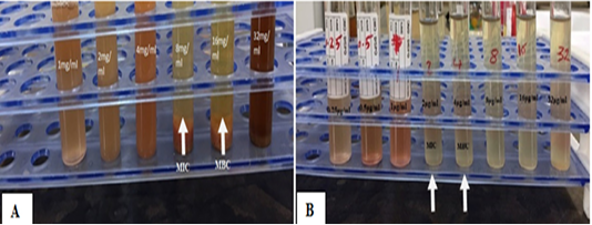
Figure 1: A MIC of C. erictus leaves extract against Staph.aureus , B: MIC of penicillin against Staph.aureus.
Clinical Examination
The result of temperature , heart rate and respiratory rate that examined during one week after skin wound infected showed there is no significant increase or decrease (p≤0.05) between different groups (T1,T2 and T3)comparing with control positive group (C),Table (2).That’s may be due to the normal phytochemical constituents mainly tannin and eugenol which present in C .erictus and clove buds (Baydaa Hameed et al., 2015) and (Shohyeb, et al., 2013).We are in agreement with (Vahid Nikouia et al., 2017) who recorded that clove oil exerts anti-inflammatory and antipyretic properties through a decrease in inflammatory cells.
Scar Formation
The amount of scar tissue take indication for quality of wound healing, the results of macroscopic evaluation at 15th day Post wound induction showed the formation of scar tissue in control group more than in other groups. While in 30th day Post wound induction the scar tissue decreased in all groups but still in control group more quantity than in other treated groups, that characterized little amount of scar tissue that may be due to mechanisms occur to different degrees during the four types of healing. Primary healing occurs when a wound is closed within hours of its creation. Delayed primary healing occurs when the wound is purposefully left open, for some interval, prior to closure. Healing by secondary intention occurs in wounds that are left to heal with or without topical therapy. Here, dressing changes are performed until the wound closes by contraction and epithelialization. Finally, partial thickness wounds or wounds involving the epidermis and part of the dermis heal by epithelialization that agreement with (Glat and Longaker, 1997; García-Alcantara et al., 2013).
Table 2: Physical observation of dogs during one week of wound.
|
Groups/ Parameters |
Temperature Mean ± S.E |
Respiratory rate Mean ± S.E |
Heart rate Mean ± S.E |
|
C (control positive) |
38.15 ± 0.31 A |
22.14±1.16 B |
95.71±5.66 C |
|
T1 (C.erictus) |
38.05 ± 0.41 A |
23.30±0.90 B |
88.31±4.96 C |
|
T2 (clove buds) |
37.83± 0.37 A |
21.28±1.52 B |
90.23±7.15 C |
| T3 (combination) | 37.47± 0.40 A | 22.37±0.97 B |
85.43±6.37 C |
-N=8 -LSD=5.8
-different capital letters denote the differences between groups.
The scar formation in T3 group (combination) was less amount when compared with T1 and T2 group that may be due to the mixture between two plants potent stimuli for initiation of an inflammatory response and thus adhesions formation is surgical trauma. Routine surgical procedures involve various degree of tissue handling that initials tissue abrasion, desiccation, ischemia, bleeding, infection and exposure to foreign materials, any of these factors can initiate inflammatory responses which eventually lead to adhesion formation (Liakakos et al., 2001; Mittal et al., 2014).
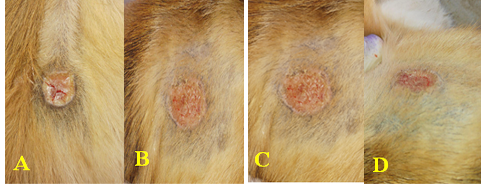
Figure 2: Scar tissue at 30th post operation A: control group B: treated one group C: treated two group D: treated three group
Histopathological Findings
The histopathological findings of the skin after inducing of wound at 3*2 cm in size were as following:
Control Group (C):
At 15th Day Post wound induction: The histopathological examination in control group at 15th day Post wound induction showed marked angiogenesis which characterized by inflammatory zone, also there were neutrophils with macrophage in dilated blood vessels and fibroblast in wound area (Figure 6).
At 30th Day Post wound induction: While, the control group at 30th Post wound induction, showed new blood vessels, granulation tissue invasion the area with presence active fibroblast (Figure 4).
Treated one Group (T1)
At 15th Day Post wound induction: The histopathological examination in treated group at 15th day Post wound induction showed there was the section showed widely scar tissue with irregular fibrosis in the wound area (Figure 3).
At 30th Day Post wound induction: Histopathological sections of treated group at 30th day Post wound induction, showed that thick scar tissue with presence the irregular connective tissue with still present inflammatory cells (Figure 5).
Treated Two Group (T2)
At 15th Day Post wound induction: The histopathological examination in treated group at 15th day Post wound induction showed there was thin scar tissue with infiltration the inflammatory cells that invaded the irregular connective tissue (Figure 6).
At 30th Day Post wound induction: Histopathological sections of treated group at 30th day Post wound induction, showed normal structure of the skin in area faraway from wound area with regular connective tissue with presence the epithelial cells with high collagen level (Figure 8).
Treated Three Group (T3)
At 15th Day Post wound induction: The histopathological examination in treated group at 15th day Post wound induction showed there was new blood vessels with presence the epithelization process with present collagen deposition in wound area (Figure 7).
At 30th Day Post wound induction: Histopathological sections of treated group at 30th day Post wound induction showed skin returned to near the normal structure which consist from normal epithelial cells with presence the blood vessels and still present inflammatory cells (Figure 9). The results of histopathological examinations of all groups at 15th day Post wound induction showed that, the treated three group (mix) had less pathological findings, and the epithelial cells in wound area showed mild pathological changes, also early and extensive regeneration of epithelial cell than control group, all these results indicated that the clove and Conocarpus erectus together support adequate nutrient for all body that included skin in addition the oxidation glucose supply brain in few second while in natural condition adequate few minutes, this observation was described by other workers (Nagamine et al., 2009; Mittal et al., 2014).
There is granulation tissue formation in the area on control group, T1 and T2 at 15 days, may indicate, improper environments in this area which need to attract fibroblasts and angioblast by growth factor which produce by microphages. Fibroblasts produce collagen fibers and angioblasts form capillary blood vessels to form granulation tissue and the degree of granulation tissue depend on degree of tissue destruction. The Presence of myofibroblasts in granulation tissue in T3 group which originated from mature fibroblasts indicated, there is sever destruction and these cells attempt to contract the affected area due to their active as smooth muscle ells. This idea was agreed with Kavi Kishor et al. (2015). In present study the wound healing appeared early and extensive in T3 group, comparing to other groups, with little scar tissue formation this is due to activation, mast cells express histamine, leukotrienes, prostanoids, proteases, and many cytokines and chemokines. These mediators may be pivotal to the genesis of an inflammatory response. By virtue of their location and mediator expression, mast cells are thought to play an active role in many conditions such as allergy, parasitic diseases, atherosclerosis, malignancy, asthma, pulmonary fibrosis, and arthritis it will be useful. With a successful trial, a treatment course is initiated (Kulinski et al., 2016). The histopathological findings at 30th day Post wound inductionappeared clear changes in quality of wound healing, that the epithelial tissue in T3 group like normal with absence of scar while in other groups still scar tissue present at 30th Post wound induction that due to Stimulates the release of vasculogenic stem cells from the bone marrow Increases vascular endothelial growth factor (VEGF) synthesis Promotes better macrophage recruitment by improving the oxygen gradient.
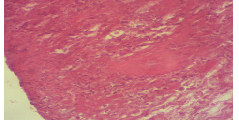
Figure 3: Photomicrography of T1 group of 15th days post operation showed scar tissue and irregular fibrosis at the wound area (H&E 100X).
The mix two plants is necessary for hydroxylation of proline and lysine, the polymerization and crosslinking of procollagen strands, collagen transport, fibroblast and endothelial cell replication, effective leukocyte killing, angiogensis and many other processes (Kavi Kishor et al., 2015).
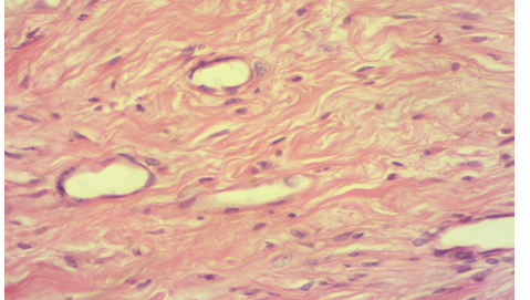
Figure 4: Photomicrography of control group at 30th days post operation showed new blood vessels, granulation of tissue invasion the area with the presence of active fibrosis (H&E 100X).
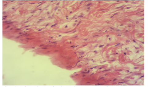
Figure 5: Photomicrography of T1 group at 30th days post operation showed thick scar tissue with presence of irregular connective tissue with inflammatory cells (H&E 100X).
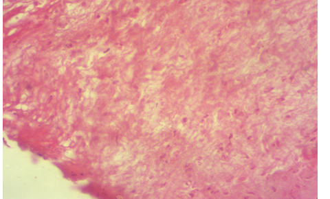
Figure 6: Photomicrography of T2 group at 15th post operation showed thin scar tissue with infiltration of inflammatory cells that invaded the connective tissue (H&E 100X).Photomicrography of T2 group at 15th post operation showed thin scar tissue with infiltration of inflammatory cells that invaded the connective tissue (H&E 100X).)
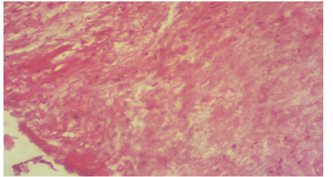
Figure 7: Photomicrography of T3 group at 15th post operation showed new blood vessels with presence of epithelization collagen deposition (H&E 100X).
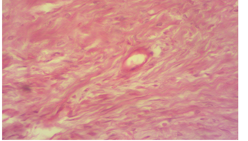
Figure 8: Photomicrography of T2 group at 30th post operation showed irregular connective tissue with presence of epithelial cell with high collagen level (H&E 100X).
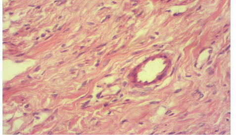
Figure 9: Photomicrography of treated three group at 30th post operation show normal epithelial cells with presence the blood vessels and still present inflammatory cells (H&E 100X).
Conclusions
From the results we aredemonstrate that clove buds and C. erictus hydro-alcoholic extracts have valuable pharmacological properties include antibacterial, anti-inflammatory and anti-pyritic in vitro and on infected wound with resistance Staph. aureus. These Features came from the constituents of plants particularly eugenol oil (clove) and tannin (C. erictus).
Acknowledgments
The authors grateful to all the Staph of Departments of Microbiology, College of Veterinary Medicine at Al-Qasim Green University for their helps and support.
Conflict of interest
There is no conflict of interest.
Authors Contribution
Both authors are involved equally in the work and writing of this article and approved it in publication.
References


