Advances in Animal and Veterinary Sciences
Research Article

Successful Treatment of Dog’s Bite Wounds in Two Sheep by a Caprine Amniotic Membrane with Long-Term Follow-Up
Karima A Al Salihi1*, Ali Abbass Ajeel1, Kasim Obaid Hussein Ali2
1College of Veterinary Medicine, Al Muthanna University, Iraq; 2College of Medicine, Al Muthanna University, Al-Hussein Teaching Hospital. Iraq.
Abstract | This is a case report describing application of Amniotic Membrane (AM) in the treatment of penetrated dog’s bite wounds in two sheep. The AM was collected from a goat during caesarean section and inserted into the bite injuries after preparation. Both cases were followed up until healing. Complete regeneration with normal histological features was evident after six weeks.An interesting effect of AM on regeneration of the bite wounds was apparent and to our knowledge, this is the first report concerning treatment of dog’s bite wounds by AM.
Keywords | Dog’s bite wounds, Amniotic membrane, Lacerated wound, Regeneration, Repair
Editor | Kuldeep Dhama, Indian Veterinary Research Institute, Uttar Pradesh, India.
Received | September 06, 2018; Accepted | December 03, 2018; Published | January 07, 2019
*Correspondence | Karima A Al-Salihi, College of Veterinary Medicine, Al Muthanna University, Iraq; Email: kama-akool18@mu.edu.iq
Citation | Al-Salihi KA, Ajeel AA, Ali KOH (2019). Successful treatment of dog’s bite wounds in two sheep by a caprine amniotic membrane with long-term follow-up. Adv. Anim. Vet. Sci. 7(3): 210-213.
DOI | http://dx.doi.org/10.17582/journal.aavs/2019/7.3.210.213
ISSN (Online) | 2307-8316; ISSN (Print) | 2309-3331
Copyright © 2019 Al Salihi and Ajeel. This is an open access article distributed under the Creative Commons Attribution License, which permits unrestricted use, distribution, and reproduction in any medium, provided the original work is properly cited.
INTRODUCTION
There is an increase in the range and extent of dog bites and predation on livestock especially the sheep worldwide (Esther et al., 2007). Currently, there is a rise in the number of stray dogs in different Iraqi governorates. The available records of dog attacks on domestic animals are incomplete and fragmented. Moreover, it is believed that every year a high number of humans and animals being attacked by dogs (Al Salihi et al., 2017), but the majority of animal cases are neither reported nor brought to the veterinary hospitals. No comprehensive reporting structure exists relating to the treatment and management of dog attacks on the domestic animal in Iraq. However, the routine management and treatment of dog bite in livestock involve washing the site of bite injuries with an antiseptic solution and topical antibiotic application accompanied by systemic antibiotic and sometimes suturing of the large macerated areas (Morales et al., 2011). Amniotic membrane has been used as a wound biological dressing for almost a century (John, 2003). Furthermore, review of the literature didn’t reveal any report of intolerance or allergy in using AM due to its low immunogenicity (Gris et al., 2002; Singh et al., 2004). In clinical practice, AM has been used to treat various types of injuries, such as venous ulcers, burns, radiation lesions and arterial ulcers (Sabella, 1913; Al Salihi et al., 2017). Likewise, AM has also been applied to cutaneous wounds and expressed its ability to accelerate wound healing (Forbes et al., 2012; Al Salihi et al., 2017). Consequently, the current study describes treatment of deep lacerated dog’s bite injuries in two sheep using goat’s amniotic membrane as transplant material.
Cases presentation
Two years old ram and four years old ewe were admitted individually to Al-Muthanna veterinary hospital at day 2 and day 7 respectively after attacking by stray dogs at midnight in January 2017. The ram was suffering from several bite injuries on different sites of its body with a large, ventro-dorsal penetrated and macerated wound accompanied by cavitations between the fat-tailed area, whereas the ewe was admitted with multiple superficial bite injuries on its dorsal surface with a deep wound on its back oozing copious quantities of yellowish pus.
After examination, the large bite wound on fat-tailed area of the ram was treated with local antibiotic, irrigated with Povidone/ Iodine antiseptic, debrided and treated by inserting pieces of AM that was collected from a caesarean section of a goat with dystocia admitted to the hospital at the same time. The AM was washed with sterile phosphate buffered saline and soaked with PBS mixed with a cocktail of antibiotics composed of 400 ml normal saline containing 1,200,000 IU benzathine penicillin and 100 ml of metronidazole. Following that, the AM pieces were inserted into the wound and the wound margins were opposed by silk sutures (3/0). Three weeks later, a distinct reduction in size of the wounds was apparent and on the fourth week, slight, fibrous connective tissue scars were still apparent at the wounded areas. Complete healing of the wounds was evidenton week 6 (Figure 1. A, B, C, D, E and F). Three months after healing the ram was slaughtered and tissue samples were collected from the fat tailed area for histological investigation using routine histological processing. Four-five µm thick tissue sections were stained with Haematoxylin and Eosin stain and examined under the light microscope and photos were captured using a digital camera. The histological sections reveal normal fat tissue (Figure 2).
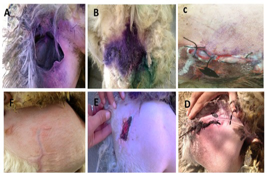
Figure 1: It shows a large, dog’s bite wound on the fat-tailed area of the ram. A&B: The ventral and dorsal surfaces of the wounded area; C: Insertion of amniotic membrane pieces into the wounded area from the ventral surface and suturing of the wound; D: Wound’s appearances on the second day after treatment revealing full closure of the wound margins; E. Reduction in size of the wound on week 4; F. Complete healing of the wound on week 6.
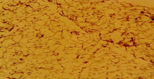
Figure 2: Microscopic view of a fat tissue section obtained from the area of the dog’s bite wound in the ram’s tail three months after healing. It shows normal morphology of the fat tissue. H & E stain, X10.
The bite injuries on the ewe’s body were cleaned from the pus, washed several times with normal saline, treated locally with antibiotic (Figure 3) and irrigated with Povidone/ Iodine antiseptic. Later on, the deep wound on the animal back was treated by insertion of AM pieces as previously mentioned in case 1. The wound was followed up and a complete healing was apparent at the end of week 5.
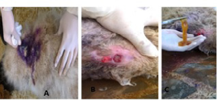
Figure 3: It shows a deep, a dog’s bite woundon the back of a 4 –years-old ewe. A. Yellow pus is evident oozing from the wound. B. Cleaning and preparation of the wound. C. Insertion of amniotic membrane pieces into the wound.
Discussion
The incidence of dogs bites and attacks on livestock has not been documented well by the concerned authorities in Iraq; in addition, no official data and no previous publications related to this matter are available. Moreover, the treatment of dogs attacks on livestock is notproperly estimated because most of the affected livestock are condemned and not used for human consumption. The dog bite wounds are usually lacerated, and their healing requires a long time and intensive care. The process of wound healing is a normal biological procedure for reparation of any tissue damage in the living body; it comprises a balanced activity of inflammatory, vascular, connective tissue and epithelial cells leading to the rebuilding of tissue integrity (Hassan et al., 2008). Following the initial clinical examination and assessment, the dog bite wounds should be assessed for the presence of infection and classified according to the type, size, and depth (Baskovich et al., 2008).
The current study dealt with the treatment of dog’s attack injuries on two sheep (2 years old ram and 4 years old ewe) which were admitted to the hospital with deep, lacerated wounds with the complication of one of the ewe’s wounds by exudation of a copious amount of yellowish pusThe basis for treatment of any lacerated wound is irrigation and removal of the necrotic tissue debris followed by cleaning (by soap) and scrubbing the wounded area to help in preventing infection because the penetrated wound injuries are difficult to be irrigated thoroughly and hence are twice as likely to become infected (Callaham, 1994; Panagiotis, 2009).
Treatment of the dog bites wounds in the present study was supported by insertion of pieces of an amniotic membrane obtained from a cesarean section of a goat with dystocia admitted to the hospital at the same time of admitting of the affected sheep. This supporting treatment resulted in complete healing of the macerated and penetrated wound in fat-tailed area in the ram and the deep, suppurative wound in the ewe. This finding, which approved the ability of caprine AMto improve and accelerte the regeneration of ovine wounds is in agreement with a similar finding reported by Subrahmanyam (1994) who approved the benefits of application of human amniotic membrane as a cover for micro skin grafts and with findings of other authors (Rejzek et al., 2001; Gajiwala et al., 2004; Baskovich et al., 2008;Rayate et al., 2016; Al Salihi et al., 2017; Susan & Blackburn, 2007) who approved the efficacy of amniotic membrane as a biological substitute to enhance the process of neovascularization and to speed the process of reepithelization in healing of skin wounds and burns.
This interesting finding of the successful supportive treatment of dog’s bite wounds by AM can be attributed to its incorporation in the wounded areas and to its effects as anti-inflammatory, anti-scarring, tissue reparation enhancement, increased deposition of extracellular matrix and promotion of fibrogenesis & angiogenesis because the fresh amniotic membrane is an active carrier for the embryonic stem cells which fix each other by desmosomes (King, 1982) and it represents a pool of cytokines and growth factors such as the epidermal growth factor, vascular endothelial growth factor, keratinocyte growth factor, fibroblast growth factor and transforming growth factor which play an essential role in wound healing (Richard et al., 2004; Rayate et al., 2016).
In conclusion, this study approved that the supportive treatment ofdog’s bit wounds by AM applicationhas an interesting effect on the healing process of such macerated, possibly infected wounds. To the authors’ knowledge, this is the first report related to the use of AM in the treatment of dog’s bite wounds cases in sheep. The authors recommend using the amniotic membrane as a supportive biological transplant in the treatment of dog bites in animals as well as human.
acknowledgements
The authors would like to thanks the owners of these animals for their Patience in the treatment of these cases.
Conflict of interest
The authors declare that there is no conflict of interest regarding the publication of this paper by any third parties.
Author’s contribution
All authors contribute equally in the clinical study, histological preparation, writing and revising the manuscript of this article.
References


