Advances in Animal and Veterinary Sciences
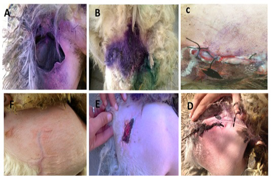
It shows a large, dog’s bite wound on the fat-tailed area of the ram. A&B: The ventral and dorsal surfaces of the wounded area; C: Insertion of amniotic membrane pieces into the wounded area from the ventral surface and suturing of the wound; D: Wound’s appearances on the second day after treatment revealing full closure of the wound margins; E. Reduction in size of the wound on week 4; F. Complete healing of the wound on week 6.
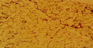
Microscopic view of a fat tissue section obtained from the area of the dog’s bite wound in the ram’s tail three months after healing. It shows normal morphology of the fat tissue. H & E stain, X10.
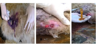
It shows a deep, a dog’s bite woundon the back of a 4 –years-old ewe. A. Yellow pus is evident oozing from the wound. B. Cleaning and preparation of the wound. C. Insertion of amniotic membrane pieces into the wound.


