Advances in Animal and Veterinary Sciences
Research Article
Adv. Anim. Vet. Sci. 4(8): 441-448
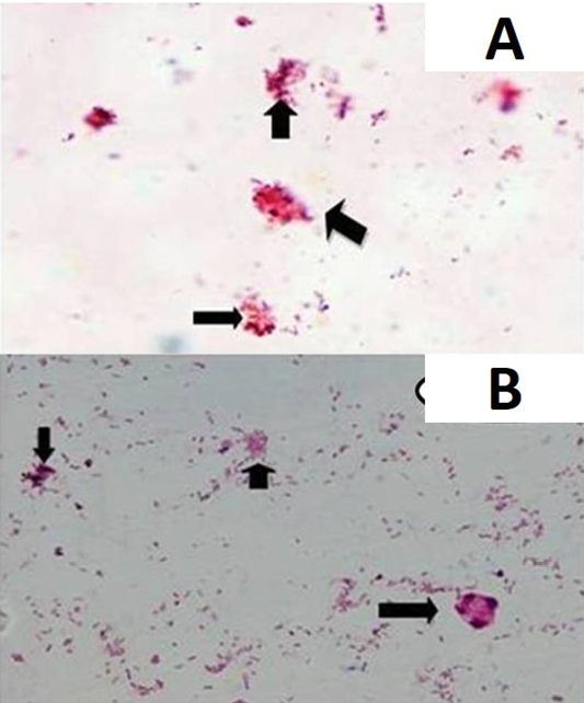
Figure 1
MAP bacilli as seen in acid fast staining in paneer samples
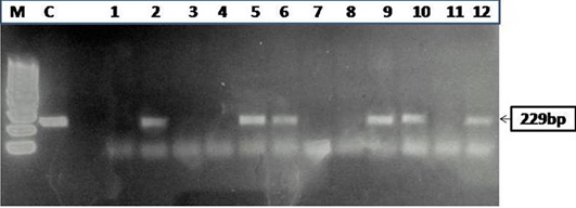
Figure 2
Agarose gel electrophoresis of PCR products obtained by IS900 PCR performed on paneer (n=55) samples
Figure 3
Dot-ELISA of paneer (n=55) samples showing brown dot for the samples positive for MAP
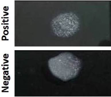
Figure 4
The presence and absence of agglutination in MAP positive and negative samples as observed by Latex Agglutination test
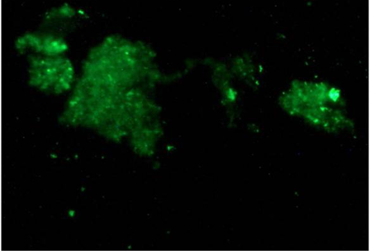
Figure 5
Green fluorescence indicating the presence of MAP bacilli in paneer samples by Indirect Fluorescent antibody test (iFAT)


