Advances in Animal and Veterinary Sciences
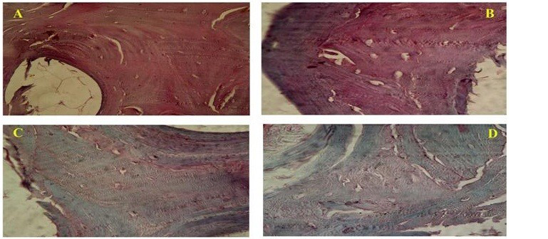
Photomicrograph showing (A-Native bone) intense GAG staining, (B-SDS treated bone) preserved GAG, (C-Acetone/Ethanol treated) less staining for GAG and (D-Freeze and Thawing group) less GAG content as compared to native bone. (Safranin-O; x40)
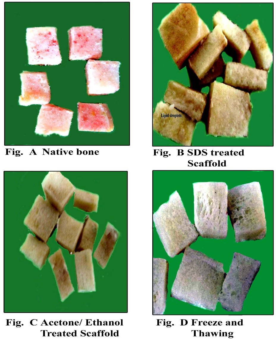
Gross appearance of the scaffolds of Native bone and decellularized bone
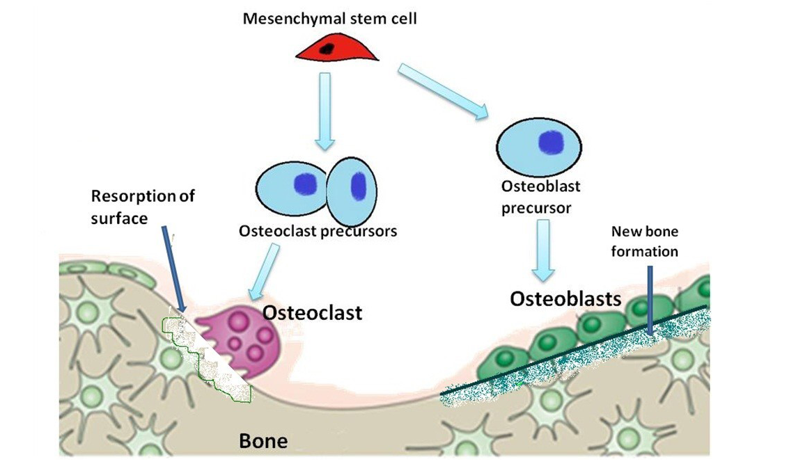
Diagram is showing evolution of osteoblasts and osteoclasts in the formation and resorption of bone
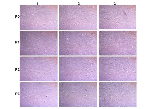
Rabbit fatal Osteoblast cell culture: P0; P1; P2 and P3 are passaged. 1; 2 and 3 images after 2; 6 and 8 days after passages. Cells become confluence (80-90% growth) within 6-8 days after passage
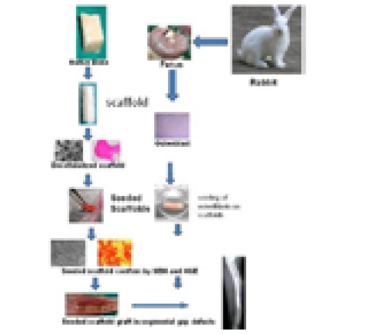
The process of graft preparation and in vitro and in vivo evaluation


