South Asian Journal of Life Sciences
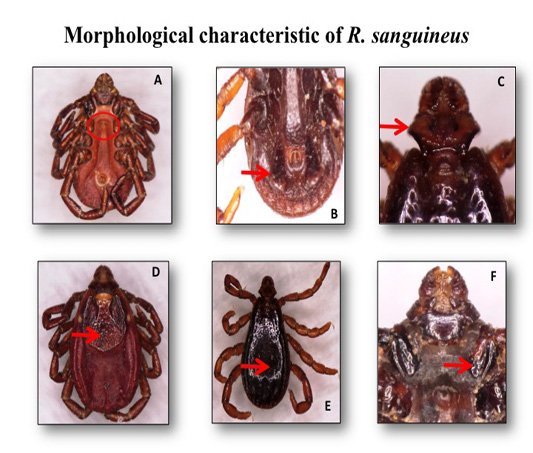
he adult female and male of the brown dog ticks Rhipicephalus sanguineus photographed by light microscope: (A) genital opening of R. sanguineus (Female); (B) adanal plate of R. sanguineus (Male); (C) hexagonal shape of basic capiculi; (D) dorsal view of R. sanguineus (Female scutum); (E) dorsal view of R. sanguineus (Male conscutum); (F)divided 1st coxa of R. sanguineus.
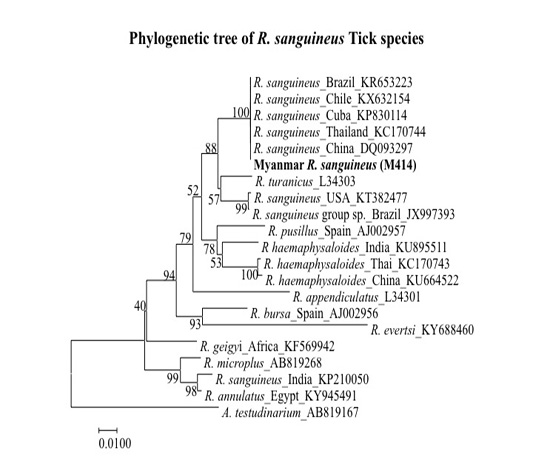
Phylogenetic tree based on R. sanguineus spp. 16S rDNA sequence. Sequences from the Rhipicephalus genera were compared with the neighbor-joining method with distance matrix calculation by Kumar-two parameters, operated by MEGA software (Version 7), using Amblyomma testudinarium as the out group. Scale bar indicates the number of mutations per sequence position. The numbers at the nodes represent the percentage of 1000 bootstrap re-samplings.


