Journal of Infection and Molecular Biology
Short Communication
Journal of Infection and Molecular Biology 2 (4): 74 – 76Microbiological Investigation of Burn Patients in Burn Intensive Units, in Quetta, Pakistan
Muhammad Irfan2, Imran Ahmed2, Muhammad Shafee1*, Abdul Malik Tareen2, Muhammad Fazal Ur Rehman2, Shabbir Ahmad Khan2
- Center for Advanced Studies in Vaccinology & Biotechnology (CASVAB) University of Balochistan, Quetta, Pakistan
- Department of Microbiology, University of Balochistan, Quetta, Pakistan
*Corresponding author:shafeedr73@gmail.com
ARTICLE CITATION:
Irfan M, Ahmed I, Shafee M, Tareen AM, Fazal Ur Rehman M, Khan SA (2014). Microbiological investigation of burn patients in burn intensive units, in Quetta, Pakistan. J. Inf. Mol. Biol. 2 (4): 74 – 77.
Received: 2014–08–22, Revised: 2014–11–08, Accepted: 2014–11–11
The electronic version of this article is the complete one and can be found online at
(
http://dx.doi.org/10.14737/jimb.2307–5465/2.4.74.76
)
which permits unrestricted use, distribution, and reproduction in any medium, provided the original work is properly cited
ABSTRACT
Thermal injury is a serious problem that requires special management of patients in burn intensive care units. This study was designed to isolate the most common pathogens and study their antibiogram profile to commonly used antibiotics. A total of 100 wound samples were collected and subjected to microbial identification. All the samples were cultured on selective medias, cetrimide and mannitol salt agar for the isolation of Pseudomonas aeruginosa and Staphylococcus aureus. After confirmation through colony characteristics and biochemical tests, all the isolates were evaluated against commonly used antibiotics viz, Amikacin, Gentamycin, Amoxicillin and Ceftazidime, for their susceptibility using disc diffusion method. P. aeruginosa (37%) was the most common pathogen followed by Staphylococcus aureus (14%) respectively. Amikacin and Ofloxacin were found with highest zone of inhibition (18 mm and 24mm) against P. aeruginosa and S. aureus. This study explores the presence of these clinically important pathogens in burn wounds.
Burn wound infections remain serious public health issue, at least in terms of morbidity and long term disability throughout the world, especially in the developing countries. Burn injuries still produce significant losses in Pakistan due to electric short circuits and misuse of gas appliances. Burns are one of the most common devastating forms of trauma and patients with serious thermal injury require immediate specialized care in order to minimize morbidity and mortality (American Burn Association, 2000). It causes mechanical disruption at the skin and also provides a suitable site for bacterial multiplication. Burn wound is richer sources of infection than surgical wounds because of the larger area involved and longer stay of patients in the hospitals (Agnihotri et al., 2004). In addition to loss of the natural cutaneous barrier to infection, coagulated protein and other microbial nutrients in the burn wound, combined with avascularity of the wound, lead to microbial colonization. The rate of nosocomial infections are higher in burn patients due to various factors such as nature of burn injury itself, immuno compromised status of the patient, invasive, diagnostic, therapeutic procedures and prolonged stay at intensive care unit (ICU) (Pruitt et al., 1998). Infection in burn patients is difficult to control due to presence of dead and denatured burn eschar and moist environment that act as a growth medium for microbes. Prolonged hospital stay and therapeutic procedures are other contributory factors that cause delay in healing and difficult to control the infection (Gang et al., 1999). Several bacterial species are common¬ly encountered in burns but Staphylococcus aureus and P. aeruginosa are the two most common gram–positive and negative pathogens, respectively (Orenstein et al., 1997; Batra, 2003). Pseudomonas is an opportunistic pathogen that colonizes in burned skin surface, produces large amounts of exopolysaccharide that binds with water and form gels and resist many commonly used antibiotics. P. aeruginosa was found as major colonizer of the burn wound because it thrives on moist burn wound surface and usually gains access to burn patients through cross contamination. It persists as a major nosocomial infection threat to burn patients. P. aeruginosa develops antimicro¬bial resistance rapidly, which complicates medical treatment of infections. P. aeruginosa is frequently isolated from patients and hospital environments and has been implicated as the cause of nosocomial infections in burn patients (Qarah et al., 2008).
Staphylococcus aureus is another most common nosocomial pathogen which has the ability to cause number of devastating complications and increasing resistance to current antibiotics (Atoyebi et al., 1992).
Therefore, this study was aimed to evaluate the most prevalent bacterial pathogen and the sensitivity of pathogens to the commonly used antibiotics.
This study was conducted in the district Quetta Balochistan, Pakistan located at 30 12’ 38’’ N 67۫ 1’8 E. It is the provincial capital with more than 2.5 million populations and is about 150 miles away from Afghan border (Hussain et al., 2012). A total of 100 wound samples were collected from 100 hospitalized patients from different hospitals (Bolan Medical College Hospital, Civil Hospital and Combined Military Hospital) in Quetta city.
All the samples were collected in sterile pre-labeled transport swabs following standard microbiology procedures. All the samples were collected from hospitalized patients in burn ICU ward at three hospitals of Quetta and were processed in the Microbiology Laboratory of Center for Advanced Studies in Vaccinology and Biotechnology (CASVAB), University of Balochistan, Quetta.
The collected swabs were inoculated onto freshly prepared selective media like Mannitol salt agar and Cetrimide agar for the isolation of S. aureus and P. aeruginosa. The plates were then incubated at 37 oC for 24 hours. Suspected isolates were presumptively identified by colony morphology, pigment formation for P. aeruginosa (pyocyanin and pyoverdine) along with biochemical tests (Catalase and Coagulase) in addition to Gram staining as described by Parvin et al. (2009).
P. aeruginosa isolates were confirmed by certain biochemical tests including citrate utilization, aesculin hydrolysis, gelatin hydrolysis, nitrate reduction and growth at 42 oC. In addition to these tests, sugar fermentation tests including glucose, sucrose, maltose was also performed. The susceptibility test of P. aeruginosa isolates were performed by Kirby bauer method (NCCLS, 1998).
The antibiogram activity against P. aeruginosa and Staph. aureus were evaluated using disc diffusion method as reported by Morteza et al. (2010). Briefly, 0.5 McFarland standardized suspension of pre-characterized clinical isolates was swabbed over the surface of Muller–Hinton agar plate. Four different antibiotics discs were placed onto the inoculated surface. After overnight incubation at 37 oC, the zone of inhibition exhibited by each antibiotic was measured (mm).
The collected was analyzed using SPSS version 16 and Chi Square test was applied with non significant difference of mean zone of inhibition of different antibiotics used against pseudomonas while the p value was < 0.05.
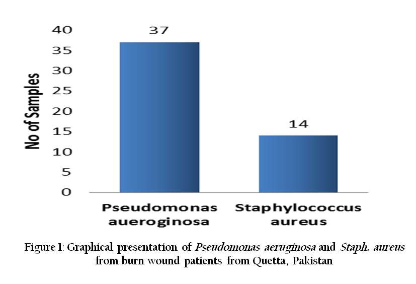
Figure 1: Graphical presentation of Pseudomonas aeruginosa and Staph. aureus from burn wound patients from Quetta, Pakistan
All the clinical isolates were collected from hospitalized patients from three different hospitals of Quetta City. Most of patients were male (60 %) and of mature and productive age group (Table 1). Most of the female patients were injured by accidental damage in kitchen while male were burnt during repair work. Out of one hundred (100) clinical isolates, 37 and 14 were identified as P. aeruginosa and Staph. aureus respectively (Figure 1). The relative proportion of pyoverdine and pyocyanin of P. aeruginosa were recorded as 49 % and 51 %, respectively. Out of the five different antibiotics used, Amikacin exhibited best zone of inhibition against P. aeruginosa with the 18 and 16 mm followed by Imipenem 15 and 13 mm, Gentamicin 13 mm for each and Ceftazidime with 11mm zone against pyocyanin and pyoverdine pigment producing P. aeruginosa. All the antibiotics were equally susceptible to both the pigments.

Out of four different antibiotics (Gentamycin, Ofloxacin, Novobiocin and Methicillin), Ofloxacin exhibited best zone of inhibition against Staph. aureus with the 24 mm followed by Novobiocin and Gentamacin with 15 and 9 mm respectively while Staphylococcus aureus were found resistant to Methicilin
Pseudomonas aeruginosa and Staphylococcus aureus are two main causes of infection in human especially in burn wounds that may be complicated further by other pyogenic organisms. Our findings are in line with (Kalantar et al., 2012: Ekrami and kalantar 2007: Nasser, 2003) who also reported the prevalence of P.aeruginosa and staph aureus 37.5 % and 20 % respectively at burn hospital in Iran. A significant number of P. aeruginosa (74%) was found in a study conducted in Tohid Burn Centre Tehran Iran (Lari et al., 1998) who suggested that this high frequency of P. aeruginosa might be due to prolonged hospital stay and intensive use of antibiotics.
Out of the five different antibiotics used, Amikacin exhibited best zone of inhibition against P. aeruginosa (18 mm and 16 mm) followed by Imipenem (15mm and 13mm), Gentamycin (13mm for each) and Ceftazidime with 11mm zone against pyocyanin and pyoverdine pigment producing P. aeruginosa (Table 2). These findings corroborate with the study by Gehan et al. (2011) who also reported that high sensitivity of Amikacin followed by Imipenem and all isolates of P. aeruginosa showed high resistance against Amoxicillin. In some studies, the Imipenem was found more sensitive against P. aeruginosa (Ronald et al., 1998; Neely and Holder, 1999).

In the present study, 93.2% P. aeruginosa isolates showed resistance against Gentamycin. This finding is similar to the study conducted in Tohid burn centre Tehran Iran, where more than 95% strains of P. aeruginosa were resistant to Gentamycin (Lari, 1998). Gentamycin is a cheap and easily available drug that is used extensively in general and hospital practice in clinically suspected Gram negative infections. This may be the main reason for the development of resistance in bacteria against this drug. This resistance may be due to many factors especially extended spectrum beta lactamase (ESBL) (Ullah et al., 2009). Out of four different antibiotics (Gentamycin, Ofloxacin, Novobiocin and Methicillin), Ofloxacin exhibited best zone of inhibition against Staphylococcus aureus with the 24mm followed by Novobiocin and Gentamycin with 15 and 9 mm, respectively. These results are in line with Ikeagwu et al. (2008) who also reported the high sensitivity of Ofloxacin against S. auerus isolates at Abakaliki, Nigeria followed by Novobiocin with 15mm, and Gentamycin with 09 mm. Staphylococcus aureus showed resistance against Methicillin (cell wall synthesis inhibitors) and our these findings are in agreement with Hafiz et al. (2002) who also reported resistance of S. aureus isolates to Methicillin at Mid–East Medical Center, Karachi, Pakistan and, also isolated it in 76 % of the samples.
P. aeruginosa is very resistant to most antibiotics and the resistance in this organism develops very rapidly. The rate of development of resistance to new antibiotics is much faster than the rate of invention and development of new antibiotics (Estahbanati, 2002). In summary, the P. aeruginosa and staph aureus are the two main sources of infection among burn patients in Quetta, Pakistan. Amikacin is the most potent drug against pseudomonas infection. The presence of these resistant strains raises great concern among patients and clinical practitioners.
ACKNOWLEDGMENT
The author acknowledges the support and technical advises of Prof Dr M Arif Awan Director CASVAB, University of Balochistan, Quetta and Prof Dr Zafar Ahmed.
REFERENCES
Agnihotri N, Gupta V, Joshi RM (2004). Aerobic bacterial isolate from burn infections and their antibiograms – A five – year study. Burns. 30: 241 – 243.
http://dx.doi.org/10.1016/j.burns.2003.11.010
PMid:15082351
American Burn Association (2000). Burn incidence and treatment in the US: fact sheet.
Atoyebi OA, Sowemimo GOA, Odugbemi T (1992). Bacterial flora of burn wounds in Lagos, Nigeria: a prospective study. Burns. 18: 448 – 451.
http://dx.doi.org/10.1016/0305-4179(92)90175-T
Batra AK (2003). Burn mortality: recent trends and socio–cultural determinants in rural India. Burns. 29: 270 – 275.
http://dx.doi.org/10.1016/S0305-4179(02)00306-6
Ekrami A, Kalantar E (2007). Analysis of the bacterial infections in burn patients at Taleghani Burn Hospital in Ahvaz, Khuzestan province. Iranin J. Clin. Infect. Dis. 2 (1): 9 – 12.
Estahbanati HK, Kashani PP, Ghanaatpisheh F (2002). Frequency of P. aeruginosa serotypes in burn wound infections and their resistance antibiotics. Burns. 28 (4): 340 – 348.
http://dx.doi.org/10.1016/S0305-4179(02)00024-4
Gang RK, Bang RL, Sanyal SC, Mokaddas E, Lari AR (1999). Pseudomonas aeruginosa septicaemia in burns. Burns. 25: 611 – 616.
http://dx.doi.org/10.1016/S0305-4179(99)00042-X
Gehan M, kamel NA, Ezzeldeen, EI–Mishad MY, Farouk RE (2011). Susceptibility pattern of P. aeruginosa against antimicrobial agents. Global Veterinaria. 6 (1): 61 – 72.
Hafiz S, Hafiz AN, Ali L (2002). Methicillin resistant Staphylococcus aureus: a multicentre study. J. Pak. Med. Assoc. 52: 312 – 315.
PMid:12481663
Hussain K, Shafee M, Khan N, Jan S, Tareen AM, Khan MA (2013). Seroprevalence of Pediatric Malaria in Quetta, Balochistan, Pakistan. Iran. J. Parasitol. 8 (2): 342 – 347.
PMid:23914251 PMCid:PMC3724163
Ikeagwu I J, Amadi ES, Iroha IR (2008). Antibiotic sensitivity pattern of Staphylococcus aureus in Abakaliki, Nigeria. Pak. J. Med. Sci. 231 – 235.
Kalantar EI, Taherzadeh S, Ghadimi T, Soheili F, Salimizand H, Hedayatnejad A (2012). Pseudomonas aeruginosa, an emerging pathogen among burn patients in Kurdistan Province, Iran. Southeast Asian J. Trop. Med. Public Health. 43(3): 712 – 717.
PMid:23077851
Lari AK, Bahrami HH, Alaghehbandan R (1998). Pseudomonas infection in Tohid Burn Centre, Iran. Burns. 24 (7): 637 – 641.
http://dx.doi.org/10.1016/S0305-4179(98)00090-4
Nasser S, Mabrouk A, Maher A (2003). Colonization of burn wounds in Ain Shams University Burn Unit. Burns. 29 (3): 229 – 233.
http://dx.doi.org/10.1016/S0305-4179(02)00285-1
NCCLs Performance of standard for antimicrobial susceptibility testing; eighth international supplement (1998). 18 (1): 100 – 513.
Neely AN, Holder IA (1999). Antimicrobial resistance. Burns. 25 (1): 17 – 24.
http://dx.doi.org/10.1016/S0305-4179(98)00134-X
Orenstein A, klein D, Kopolovic J, Winkler E, Malik Z, keller N, Nitzan Y (1997). The use of Porphyrins for eradication of Staphylococcus aureus in burn wounds infections. FEMS Immunol. Med. Microbiol. (4): 307 – 314.
http://dx.doi.org/10.1016/S0928-8244(97)00097-7
Parvin H, Motamedifar M, Nahal H (2009). Prevalent bacterial infections in Intensive Care Units of Shiraz University of medical sciences Teaching hospitals, shiraz, Iran. Jpn. J. Infect. Dis. 62: 249 – 253.
Pruitt Jr BA, McManus AT, Kim SH, Goodwin CW (1998) .Burn wound infections: current status. World J. Surg. 22: 135 – 145.
http://dx.doi.org/10.1007/s002689900361
PMid:9451928
Qarah S, Cunha AB and Dua P (2008). Pseudomonas aeruginosa infections. Available from: http://www.emedicine.com/med/topic1943.html
Ronald NJ, Pfaller MA, Doern GV, Erwin ME, Hollis RJ (1998). Antimicrobial activity and spectrum investigation of eight broad spectrum beta lactam Drugs: A 1997 Surveillance Trial in 102 Medical Centres in the United States. Diagnostic Microbiol. Infect. Dis. 30 (3): 215 – 228.
http://dx.doi.org/10.1016/S0732-8893(97)00234-4
Ullah F, Malik SA, Ahmed J (2009). Antimicrobial susceptibility and ESBL prevalence in Pseudomonas aeruginosa isolated from burn patients in the North West of Pakistan. Burns. 35: 1020 – 1025.
http://dx.doi.org/10.1016/j.burns.2009.01.005
PMid:19501980


