Journal of Infection and Molecular Biology
Short Communication
Journal of Infection and Molecular Biology 2 (3): 43 – 48Prevalence and Clinico–Pathological Features of Peste des Petits Ruminants in Different Breeds of Goats and their Response to Antimicrobials
Kamrul Islam1*, Abdul Ahad1, Altaf Mahmood2, Muhammad Mizanur Rahman3, Muhammad Zohorul Islam1, Muhammad Hazzaz Bin Kabir6, Mukti Barua4, Sharmin Chowdhury5, Muhammad Kamal Nasir2, Paritosh Kumar Biswas1
- Department of Microbiology, Faculty of Veterinary Medicine, Chittagong Veterinary and Animal Sciences University, Chittagong, Bangladesh
- Livestock and Dairy Development Department, Government of Punjab, Pakistan
- Department of Medicine and Surgery, Faculty of Veterinary Medicine, Chittagong Veterinary and Animal Sciences University, Chittagong, Bangladesh
- Department of Animal Science and Animal Nutrition, Faculty of Veterinary Medicine, Chittagong Veterinary and Animal Sciences University, Chittagong, Bangladesh
- Department of Pathology and parasitology, Faculty of Veterinary Medicine, Chittagong Veterinary and Animal Sciences University, Chittagong, Bangladesh
- Department of Microbiology & Parasitology, Faculty of Animal Science and Veterinary Medicine, Sher–e–Bangla Agricultural University, Dhaka
*Corresponding author:kamruldvm13@gmail.com
ARTICLE CITATION:
Islam K, Ahad A, Mahmood A, Rahman MM, Islam MZ, Kabir MHB, Barua M, Chowdhury S, Nasir MK, Biswas PK (2014). Prevalence and Clinico–Pathological Features of Peste des Petits Ruminants in Different Breeds of Goats and their Response to Antimicrobial. J. Inf. Mol. Biol. 2 (3): 43 – 48.
Received: 2014–05–13, Revised: 2014–06–13, Accepted: 2014–06–14
The electronic version of this article is the complete one and can be found online at
(
Http://dx.doi.org/10.14737/jimb.2307-5465/2.3.43.48
)
which permits unrestricted use, distribution, and reproduction in any medium, provided the original work is properly cited
ABSTRACT
Peste des petits ruminant (PPR) is an acute febrile viral disease of small ruminants characterized by mucopurulent nasal and ocular discharges, necrotizing and erosive stomatitis, enteritis and pneumonia. Present study was carried out on one hundred and eighty two goats during July to December, 2012. Black Bengal goats depicted higher prevalence (P = 0.66) than Crossbred and Jamunapari goats. Clinical examination revealed significantly increased heart (P = 0.00), temperature (P = 0.00) and respiratory rates (P = 0.00) in PPR affected goats (n = 87) than healthy goats (P < 0.05). Significantly higher prevalence (P < 0.00) of PPR (65%) was recorded in non vaccinated group (n = 102, P < 0.05) whereas non-significant difference in PPR was recorded with respect to breed (P = 0.66), sex (P = 0.68) and month (P = 0.50) of disease occurrence (P > 0.05). Blood samples of PPR affected goats (n = 10) exhibited significantly decreased (P = 0.02) total leukocyte count (9.31±0.53 thousand/mm3) and (P = 0.01) lymphocytes (41.20±2.26%) (P < 0.05), whereas significantly increased (P = 0.02) neutrophils (41.30±2.32%) as compared to healthy goats (n = 10) (P < 0.05). Postmortem examination of PPR suspected goats revealed characteristics zebra stripe lesions at ileo–caecal junction. The study suggests that PPR can affect goat population irrespective of age, breed and sex. Supportive therapy with Oxytetracycline can enhance recovery of the PPR affected goats and vaccination can reduce the disease prevalence leading to effective control and prevention.
Peste des petits ruminant is an acute and highly contagious viral disease of small ruminants caused by a non–segmented negative strand RNA virus. International organizations for animal health (OIE) have identified PPR as a notifiable and economically important transboundary viral disease of small ruminants associated with high morbidity and mortality. PPR was first reported in the Ivory Coast of West Africa and was later found in other parts of the world including sub–Saharan Africa, the Arabian Peninsula, the Middle East, and the parts of Asia (Balamurugan et al., 2012). PPR is one of the most widespread, infectious and contagious disease which drastically affect different breeds of goats of Bangladesh. Black Bengal goat is more sensitive to PPR than others and the rate of incidences is highest during the rainy season and in the dry agro–climatic zones (Hegde et al., 2009). Virus shedding occurs in exhaled air, in secretions and excretions from the mouth, eye and nose, and in feces, semen, and urine (Maganga et al., 2013). Clinical pictures of PPR showed sudden onset of depression, high rise of temperature, mucopurulent discharges from the eyes and nose, sores in the mouth, difficult breathing and cough, foul–smelling diarrhea (Rahman et al., 2011). In Bangladesh a live attenuated conventional PPR vaccine was developed by Bangladesh Livestock Research Institute (BLRI) and currently being used in the country (Rahman et al., 2011). This vaccine is highly effective in control against PPR in goats.
A total of one hundred and eighty two goats were registered at the Upazila Veterinary Hospital, Cox’s Bazar, Bangladesh during July, to December, 2012 and selected as study population. Diagnosis was made by means of anamnesis, clinical signs, blood parameters and by postmortem features. A PPR case was initially suspected through close inspection of signs of pneumo–enteritis complex. Blood samples were randomly collected from jugular veins of clinically affected (n = 10) and healthy goats (n = 10) with sterile syringe into the tubes containing EDTA @ 2mg/mL for hematological studies. Blood samples were analyzed for determination of hemoglobin, packed cell volume, total erythrocyte count, total leukocyte count and differential leukocyte count using light compound microscope blood examination technique (Tibbo et al., 2004). Postmortem examination was carried out in two PPR suspected dead animals. A PPR case was tentatively diagnosed if postmortem examinations reveal the presence of erosive and hemorrhagic ulcerative lesions in all organs of the digestive system from the mouth to the colon, characteristics Zebra stripe in the mucosa of colon, enlarged lymphnodes, congested liver, necrosis and hemorrhagic plugs in the cecum (Sahinduran et al., 2012). /p>
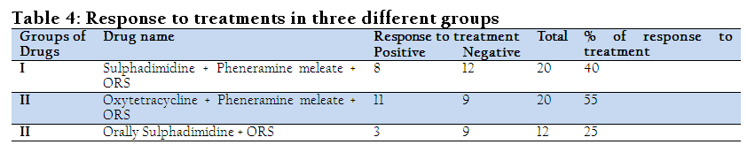
Three different treatment regimens were conducted in PPR affected goats for evaluating the treatment. Group I: Animals were administered with Sulphadimidine @ 165 mg/Kg on first day and 82.5 mg/Kg for the next four days, Pheneramine meleate @ 1mg/Kg and an oral rehydration saline (ORS) @ 1.25gm/L of water/goat for five days; Group II: were administered with Oxytetracycline–100mg @ 10mg/Kg body weight for five days, Pheneramine meleate @ 1mg/Kg and an oral rehydration saline (ORS) @ 1.25gm/L of water/goat for five days; and Group III: were treated with oral Sulphadimidine @ 200mg/Kg on first day and 100mg/Kg for the next four days and an oral rehydration saline (ORS) @ 1.25gm/L of water/goat for five days; A diluted solution of Potassium permanganate (0.5%) and glycerin was smeared on the gums, tongue and palate (oral cavity) in all three PPR affected groups for soothing effect ( Narayanan et al., 2008).
All the data including categorical variables and continuous variables from the goats clinically examined were entered into MS excel (Microsoft office excel–2007, USA). Data management and data analysis were done by STATA version–12.1 (STATA Corporation, College Station, Texus). Prevalence was calculated according to different categories of the explanatory variables. Descriptive analysis was done by means of creating histogram and boxplot. To identify the association between a categorical explanatory variable with the outcome (occurrence of PPR), chi– square (χ2 test) test was performed and for continuous variables t–test was conducted to evaluate if the mean values between positive and negative group of animals differed significantly or not. An association was regarded as significant if the p ≤ 0.05.
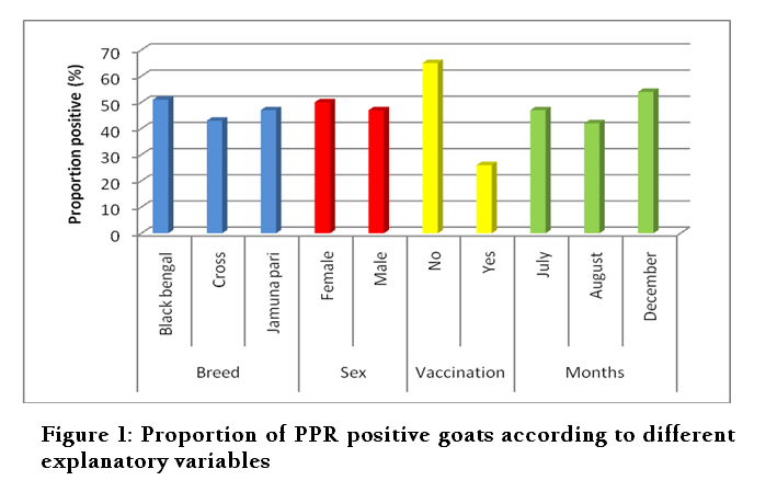
The proportions of PPR cases according to different explanatory variables are shown in Figure 1. The overall prevalence of PPR during the study period was estimated to be 47% (95% CI 34.6–59.4%). Prevalence of PPR in black Bengal goats were 51% (95% CI 39.9–62.0%). On the other hand, 43% (95% CI 28.0–57.9%) and 47% (95% CI 34.6–59.4%) were recorded in Crossbred and Jamunapari goats, respectively. Between male and female goats, the proportion of positive goats were nearly similar 47% (95% CI 38.2–55.8%) and 50% (95% CI 37.1–62.9%), respectively. Proportion of PPR positive goats were varied remarkably according to vaccination status. Twenty six percent (95% CI 16.4–35.6%) goats which were vaccinated and 65% (95% CI 55.7–74.3) among non vaccinated goats were positive to PPR. Seasonal variation was not very notable in this present study. Boxplot (Figure 2) is presented with the median, minimum, maximum, 25th and 75th percentile values of age between the 2 groups of goats (PPR negative and positive) investigated the median age of PPR– positive goats was comparatively higher than PPR– negative goats. Boxplot (Figure 3) is showed with the median, minimum, maximum, 25th and 75th percentile values of temperature between PPR negative and positive goats. The median temperature was higher in PPR positive goats than the PPR– negative ones. Boxplot (Figure 4) is portrayed with a boxplot showing the median, minimum, maximum, 25th and 75th percentile values of heart rate between PPR negative and positive goats, respectively.
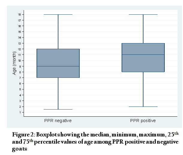
Figure 2: Boxplot showing the median, minimum, maximum, 25th and 75th percentile values of age among PPR positive and negative goats
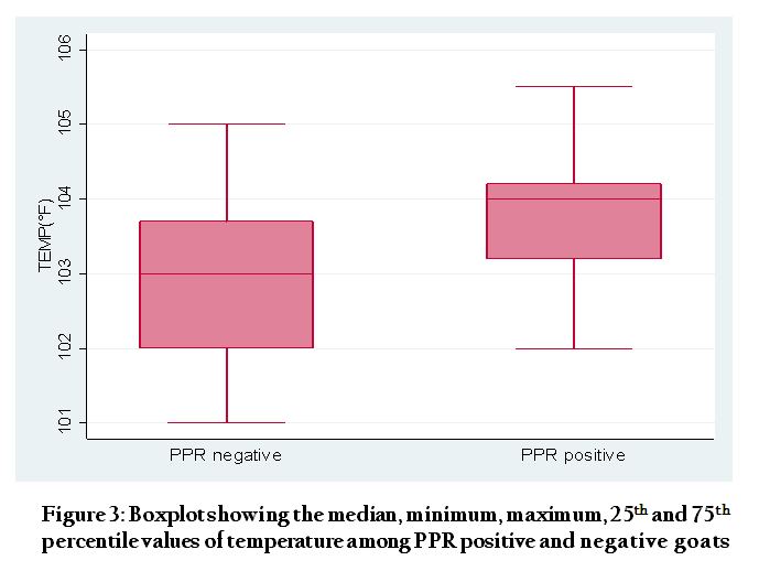
Figure 3: Boxplot showing the median, minimum, maximum, 25th and 75th percentile values of temperature among PPR positive and negative goats
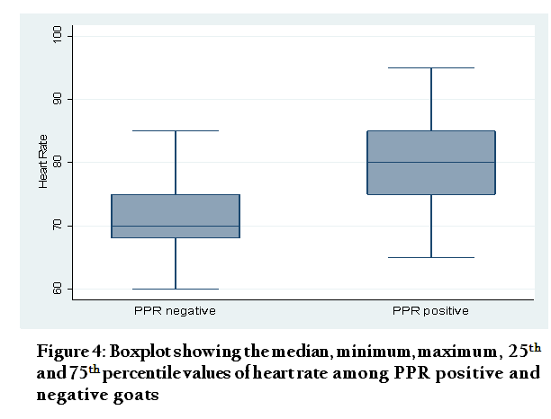
Figure 4: Boxplot showing the median, minimum, maximum, 25th and 75th percentile values of heart rate among PPR positive and negative goats
The median heart rate was observed in PPR–positive goats was higher compared with the negative goats. Boxplot (Figure 5) is presented with the median, minimum, maximum, 25th and 75th percentile values of respiratory rate between PPR negative and positive goats respectively. The median respiratory rate was higher in PPR positive goats compared with negative goats. Table 1 presented with the statistics of 182 goats according to different categorical variables. The prevalence of PPR was evenly distributed in Black Bengal, Crossbred and Jamuna Pari goats (P = 0.66). Its occurrence was proportionately but not significantly higher in female goats (p = 0.68). The rate of PPR in the non–vaccinated goats was 65% (95% CI 55.7–74.3) significantly higher compared with the vaccinated goats, 26% (95% CI 16.4–35.6%) (P = 0.00). According to season, the prevalence was recorded higher in the winter month (December) than in the rainy ones (July and August) (P = 0.50). During July, August and December the prevalence of PPR were 47%, 42% and 54%, respectively, and there were no significant difference (P > 0.50) in its occurrence in the said three months of investigation. Comparative mean values of different continuous variables such as age (in month), temperature, heart rate, respiratory rate etc were shown in Table 2. In the PPR positive goats the mean age was 10.20± 0.41, 9.39±0.40 months in the negative goats (P = 0.16). In the PPR affected goats the mean temperature, heart rate and respiratory rate – all were significantly higher (P = 0.00) compared with PPR–negative goats.
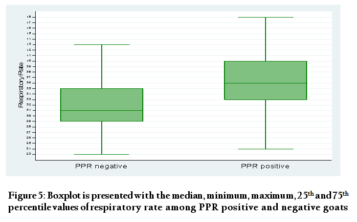
Figure 5: Boxplot is presented with the median, minimum, maximum, 25th and 75th percentile values of respiratory rate among PPR positive and negative goats
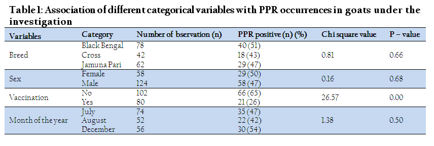
Table 1: Association of different categorical variables with PPR occurrences in goats under the investigation
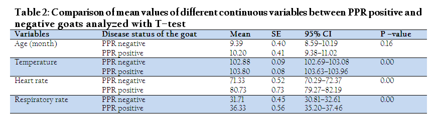
Table 2: Comparison of mean values of different continuous variables between PPR positive and negative goats analyzed with T–test
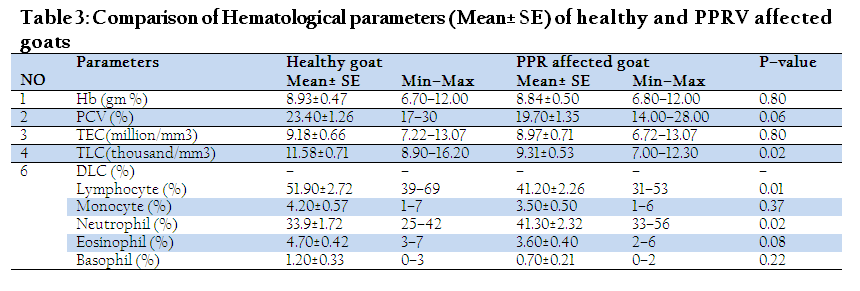
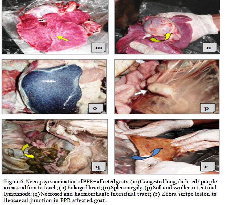
Hematological pictures of 10 PPR affected goats compared with 10 normal goats shown in Table 3. In the diseased goats the Mean ± SE values of Hb (8.84±0.50 gm %), PCV (19.70±1.35), TEC (8.97±0.71 million/mm3), TLC (9.31±0.53 thousand/mm3), Lymphocyte (41.20±2.26 %), Monocyte (3.50±0.50 %), Neutrophil (41.30±2.32%), Eosinophil (3.60±0.40%), Basophil (0.70±0.21%) all were lower compared with the healthy goats. The recovery rates in the three treatment groups are presented in Table 4. Treatment with Oxytetracycline had higher 55% (95% CI), recovery in comparison to two other groups. At necropsy, severe dehydration, dirty white, pseudo membranes, erosions on the gums, soft and hard palates, tongue and cheeks, consolidated lungs, necrosed and congested liver, enlarged heart, splenomegaly, soft and swollen intestinal lymphnodes, zebra stripe lesion in ileo–caecal junction etc were recorded in Figure 6.
PPR is one of the most commonly occurred diseases in Bangladesh. In the present study higher prevalence of PPR was found in Black Bengal goat than Jamuna Pari and Crossbred goats. This is similar to the finding published previously by Sarker and Islam (2011). The prevalence of PPR was slightly higher in male goats compared to females which in contrast to the findings reported by Abdalla et al. (2012). It is not clear whether PPR has any sex predilection which needs further investigation to confirm. The prevalence of PPR in vaccinated goats was lower compared with non–vaccinated goats. This finding is similar with an agreement previously published by Islam et al. (2012). The reason for higher prevalence of PPR in non vaccinated animals may be due to absence of antibodies to PPRV reported by Banik et al. (2008). The disease occurrence was highest during the month of December, and lowest in the month of August. Based on season, apparently the disease does not differ but during winter (December) the percentage of occurrence was comparatively higher than in rainy season which is similar to the finding previously reported by Gupta et al. (2007). Apparently the disease does not differ between different age groups, but median age was comparatively higher in PPR–positive goats than negative. It was previously reported that the goats of age between 4 to 12 months were more prone to PPR than older (>1 year) Gupta et al. (2007). The temperature was significantly higher in PPR positive goats than negative goats. Similar higher temperature was reported by Khan et al. (2005). The present study also recorded a higher heart rate and respiratory rate in PPR–positive goats than negative. This may be the results of severity of dehydration and nutritional status of the PPR–positive animals. On the other hand respiratory rate might have increased in the PPR affected goats because of pneumonic condition. Similar high respiratory rate in PPR–goats was reported by Radostits et al. (2000).
Hematological parameters were lower in the PPR–goats compared to healthy goats, except neutrophil counts. Total erythrocyte counts (million/mm3) and level of Hb (gm %) were similar in both the PPRV–infected and healthy goats and PCV (%) was lower. The Hb (gm %) and PCV (%) reflect the downturn of the TEC counts. Total leukocyte counts were higher in healthy goat as compared to affected goats though this value does not cross the normal limit. The counts for neutrophils were higher in the PPRV–affected goats whereas lymphocyte counts in the PPRV–affected goats were lower than healthy goats. Monocyte, Eosinophil and Basophil counts were lower in the PPRV–affected goats than in healthy goats. Postmortem examination of two dead animals reveals erosions on the gums, soft and hard palates, tongue and cheeks and into the esophagus. Erosive and hemorrhagic ulcerative lesions were located in all organs of the digestive system from the mouth to the colon, consolidated and pneumonic lungs, characteristic Zebra striping in the mucosa of colon etc. Similar findings were previously published by Sahinduran et al. (2012). The success rate of treatment with parenteral (I/M) administration of Oxytetracycline was higher (55%) than parenteral (I/M) use of sulphadimidine (40%) and oral/gut acting sulphadimidine (25%), supported by an agreement with Gupta et al. (2007).
It was concluded that PPR can infect goat population irrespective of age, breed and sex. Vaccination and supportive therapy of Oxytetracycline can significantly decrease its rate of occurrences leading to effective control and prevention.
ACKNOWLEDGEMENTS
Authors are grateful to the Department of Microbiology, Faculty of Veterinary Medicine, Chittagong Veterinary and Animal Sciences University, Bangladesh for giving laboratory support to the present research study and also to the Upazila Veterinary Hospital, Cox’s Bazar sadar, Cox’s Bazar for sampling assistances.
REFERENCES
Abdalla SA, Majok AA, Malik KHEI, Ali AS (2012). Sero–prevalence of peste des petits ruminants virus (PPRV) in small ruminants in Blue Nile, Gadaref and North Kordofan States of Sudan. J. Public Health Epidemiol. 4(3): 59 – 64.
Balamurugan V, Saravanan P, Sen A, Rajak KK, Venkatesan G, Krishnamoorthy P, Bhanuprakash V, Singh RK (2012). Prevalence of peste des petits ruminants among sheep and goats in India. J. Vet. Sci. 13(3): 279 – 285.
http://dx.doi.org/10.4142/jvs.2012.13.3.279
PMid:23000584 PMCid:PMC3467403
Banik SC, Podder SC, Samad MA, Islam MT (2008). Sero surveillance and immunization in sheep and goats against Peste des Petits Ruminants in Bangladesh. Bangl. J. Vet. Med. 6(2): 185 – 190.
Gupta SD, Biswas PK, Habib S, Debnath NC (2007). Prevalence of PPR in the greater Chittagong district of Bangladesh. Sci. 3 (2): 36 – 41.
Hegde R, Gomes AR, Muniyellappa HK, Byregowda SM, Giridhar P, Renukaprasad C (2009). A short note on peste des petits ruminants in Karnataka, India. Rev. Sci. Tech. 28(3): 1031 – 5.
PMid:20462160
Islam MA, Khan MSI, Kader HA, Begum MR, Asgar MA (2012). Prevalence of PPR of Goat and Their Response to Antibiotic Treatment at Mirzaganj Upazila of Patuakhali District. J. Environ. Sci. And Natural Resources, 5(2): 181 – 184.
Khan MR, Haider MG, Alam KJ, Hossain MG, Chowdhury SMZH, Hossain MM (2005). Pathological investigation of Peste des Petits Ruminants (PPR) in goats. Bang. J. Vet. Med. 3(2): 134 – 138.
Maganga GD, Verrier D, Zerbinati RM, Drosten C, Drexler JF, Leroy EM (2013). Molecular typing of PPRV strains detected during an outbreak in sheep and goats in south–eastern Gabon in 2011, http://www.virologyj.com/content/10/1/82, Virol. J. 10: 82.
Narayanan R, Gopu P, Baegan S, Barathidasan (2008). Clinical Management in an outbreak of Peste Des Petits Ruminants in Barbari Goats. Vet. World. 1(3): 81 – 82.
Radostits OM, Gay CC, Blood DC, Hinchcliff KW (2000). Diseases caused by viruses and Chlamydia. In: Veterinary Medicine, 9th ed., W. B. Saunders Company Ltd., London, 1077 – 1079.
Rahman MA, Shadmin I, Noor M, Parvin R, Chowdhury EH, Islam MR (2011). Peste des petits ruminants virus infection of goats in Bangladesh: Pathological investigation, molecular detection and isolation of the virus. The Bang. Vet. 28(1): 1 – 7.
Sahinduran S, Albay MK, Sezer K, Ozmen O, Mamak N, Haligur M, Karakurum, Yildiz R (2012). Coagulation profile, hematological and biochemical changes in kids naturally infected with peste des petits ruminants. Trop. Anim. Health. Prod. 44: 453 – 457.
http://dx.doi.org/10.1007/s11250-011-9917-y
PMid:21732067
Sarker S, Islam MH (2011). Prevalence and Risk Factor Assessment of Peste des petits ruminants in Goats in Rajshahi, Bangladesh. Vet. World. 4(12): 546 – 549.
http://dx.doi.org/10.5455/vetworld.2011.546-549
Tibbo M, Jibril Y, Woldemeskel M, Dawo F, Aragaw K, Rege JEO (2004). Factors Affecting Hematological Profiles in Three Ethiopian Indigenous Goat Breeds. Intern. J. Appl. Res. Vet. Med. 2(4): 300.


