Journal of Infection and Molecular Biology
Short Communication
Journal of Infection and Molecular Biology 2 (1): 16 – 18Seroprevalence of Toxoplasma gondii Infection in Camels (Camelus dromedaries) in and around Bahawalpur Region of Pakistan
Umer Naveed Chaudhry1*, Asad Amanat Ali2, Shoaib Ashraf1, Muhammad Tanveer Khan1, Syed Maaz Nadeem3, Kamran Ashraf1
- Department of Parasitology, University of Veterinary and Animal Sciences, Lahore, Pakistan;
- Poultry Research Institute Rawalpindi, Pakistan; Department of Pathology, University of Veterinary and Animal Sciences, Lahore, Pakistan
*Corresponding author: unchaudh@ucalgary.ca
ARTICLE CITATION:
Chaudhry UN, Ali AA, Ashraf S, Khan T, SM Nadeem, Ashraf K (2014). Seroprevalence of Toxoplasma gondii infection in camels (Camelus dromedarius) in and around Bahawalpur region of Pakistan. J. Inf. Mol. Biol. 2 (1): 16 – 18.
Received: 2013–07–22, Revised: 2013–12–30, Accepted: 2014–01–04
The electronic version of this article is the complete one and can be found online at
(
http://dx.doi.org/10.14737/jimb.2307-5465/2.1.16.18
)
which permits unrestricted use, distribution, and reproduction in any medium, provided the original work is properly cited
ABSTRACT
Toxoplasma gondii is an intracellular parasite, which infects human and animals by ingestion of tissue cyst, raw or undercooked meat or oocyst from soil, vegetables, fruits, water, soil and food contaminated by cat faeces or by transmission through the placenta, milk and blood transfusion. Seropositivity levels vary widely among different regions of the globe and according to sociocultural habits, geographic factors; climate and transmission routes and typically rise with age. In view of the worldwide importance of T. gondii, a study was conducted to determine the prevalence of T. gondii antibody in camels by using Toxoplasma Latex Test Kit. The overall prevalence of T. gondii infection in camels was recorded as 10%. Two camels were found seropositive at 1:16 dilution showing residual or nonspecific immunity, five camels were found seropositive at 1:128 showing acquired or evolving immunity, whereas three camels were positive at antibody titre of 1:256 giving an evidence of present infection. It was also noted that seropositivity of T. gondii in camels was higher in age group from 6–10 years; infection was higher in female camels having abortion history.
Toxoplasma gondii is an intracellular protozoan parasite (Smith, 1995) which infects humans as well as wide variety of mammals and birds (Hill et al., 2005). Toxoplasmosis is found throughout the world and tends to be more prevalent in tropical climates (Dubey, 1999). The organism was first discovered by Nicolle and Manceaux (1908) as a tissue parasite of gondii (an African rodent), and Darling found it in Man (Subash, 1990). The infection has been confirmed in some 200 species of mammals including man and in domestic / wild felines, which are the only definitive hosts (Pedro et al., 2003).
The source of transmission is the ingestion of vegetables, fruits, water, soil, food contaminated by cat faeces, raw or undercooked meat. Flies and cockroaches may act as a mechanical carrier to transfer oocysts to different varieties of foods. Other sources include transplacental transmission, from mother to the offspring through milk, transplantation of organs, transfusion of blood and venereal transmission (Pedro et al., 2003).
T. gondii can cause severe acquired infection in animals and human beings, which may be localized or generalized. Lymphadenitis (deep cervical nodes) is the most frequently observed clinical sign. Other signs include fever, retinochoroiditis, uveitis, malaise, muscle pain, muscle fatigue, sore throat, headache, hepatitis, myocarditis and pneumonia. Encephalitis is an important sign of Toxoplasma in later stages. During the 1980,s Toxoplasmic encephalitis in humans emerged as a common complication associated with AIDS (Subash, 1990).
As far as congenital infection is concerned, animals and pregnant women develop the most serious side effects leading to spontaneous abortion, still birth, birth defects, mummification, neonatal losses or fetal abnormalities including microcephalya, hydrocephalya, brain calcifications, psychomotor & mental retardation. The mechanism of vertical transmission is not yet understood (Remington et al., 1995).
Depending upon the geographic location, disease has zoonotic importance in human population. In human 15 – 80 % population is infected with toxoplasmosis. Approximately 500 million populations are estimated to have antibodies of T. gondii infection (Subash, 1990). Study has shown that between 16% to 40% of the human population in North America and Great Britain, 50% to 80% of the populations in Europe and Latin America have antibodies of T. gondii, indicating that they have got infection at some time (Pedro et al., 2003).
Serodiagnosis has been a reliable tool to diagnose Toxoplasma infection in both human and animals, using various serological tests, such as indirect haemagglutination, indirect immuneflorescent technique, and Enzyme linked immunosorbant assay and latex agglutination test (Ahmed et al., 1983). Due to increasing risk of public health by ingestion of contaminated meat, toxoplasmosis has becomes extremely important zoonotic disease.
Camel meat is commonly being consumed, and is the most vulnerable to the exposure of toxoplasmosis which may become the potential source of infection for the consumers; so for as no literature could be traced relating to the investigation of toxoplasmosis in Pakistan in camels. Therefore, keeping in view the importance of disease, study on seroprevalence of T. gondii in camels was carried out, which would be helpful to adopt the control measures against the diseases in humans.
A total of 100 blood samples of camels were collected at random from various camel colonies of Bahawalpur. The record/history of each animal was recorded in performa. Under aseptic measures, 5–10 mL of blood was drawn from each camel by vein puncture with the help of disposable syringes and was transferred to screw capped sterile test tube, slowly to avoid haemolysis. All the blood samples were labelled with number and date of collection. The samples were left for about an hour for blood clotting to occur. The clotted blood was then separated with the help of a fine loop and blood samples were centrifuged at 3500 rpm for at least 5 minutes. The supernatant sterile serum was aspirated with a pasture pipette and transferred into a screw capped vial which was stored at –20°C degree until processed for analysis.
All the serum samples were analyzed for Toxoplasma specific IgG antibodies using Latex Agglutination Test (LAT). For this purpose, the commercial Toxoplasma Latex Test Kit was used and interpreted as per manufacturer’s instruction (Novamed, Ltd.).
A total 100 blood samples of camels were collected and analysed for anti–Toxoplasma antibodies at screening dilution of 1:16, 1:128, 1:256 by using commercially available Toxoplasma Latex Kit on the principle of Latex Agglutination Test (LAT).
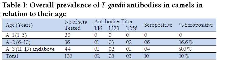
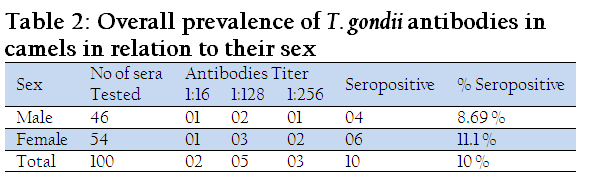
The age of camels ranged from 1–15 years and above. Blood samples were taken and divided in to 3 categories i.e. A–1, A–2, and A–3. The age categories (A–2) that ranged from 6–10 year had the highest seropositive percentage that was 16.6 % followed by A–3 (11–15 yr and above) that was 9.0 %, whereas the number of samples tested in A–1 (1–5 yr) had no positive case (Table 1). As far as the sexes of camels were concerned, 46 were male and 54 were female. Female camels have higher seropositive percentage (11.1 %) than male (8.69 %) (Table 2). The overall seropositive percentage was 10%. According to antibodies titre, 2 camels showed antibody titre at screening dilution of 1:16, 5 camels showed antibody titre at 1:128 and 3 showed antibody titre at screening dilution of 1:256 (Table 1).
Toxoplasmosis is one of the most common infections in human and animals cause by protozoan parasite T. gondii, which is responsible for significantly higher morbidity, and mortality in both human and other warm–blooded animals. Toxoplasmosis has worldwide distribution, zoonotic in nature and depending upon the geographic location 15–85% of the population can be symptomatically infected (Subash, 1990). Toxoplasmosis is also responsible for abortion and congenital defects in human and domestic livestock including sheep, goats, camels, cow and buffalo (Pedro et al., 2003).
T. gondii can cause severe acquired and congenital infection in animals and human beings associated with fever, lymphadenitis, uveitis, muscle fatigue, hepatitis, encephalitis and abortion. Serological surveys indicate that about 80% of all primary infections are asymptomatic, due to the immune system effectiveness, but variable levels of the disease can affect immunocompromised individuals (Cantos et al., 2000). The acquired immune deficiency syndrome (AIDS) has created an expanding population of susceptible individuals. Usually people suffering from both AIDS and Toxoplasmosis have been exposed to the Toxoplasma parasite earlier in life and the HIV infection simply allowed the Toxoplasma parasite to grow unchecked. The concomitant occurrences should be considered by public health policies especially in those countries with high Toxoplasma prevalence, where AIDS is concurrent with economic and public health problems (Passos et al., 2000). T. gondii infection is embrotoxic in humans. It is mainly transmitted through raw or undercooked meat and ingestion of oocytes in cat faeces (Cantos et al., 2000).
Toxoplasmosis in camels is important because of its zoonotic importance and camels are main source of meat consumption in Pakistan. Due to the zoonotic importance of Toxoplasmosis, the present study was conducted to sort out the seroprevalence of T. gondii infection in camels in & around Bahawalpur areas by using commercially available Toxoplasma Latex Agglutination Kit (LAT).
Among 100 camels examined in the present study, 2 gave an antibody titre of 1:16 which indicated residual or nonspecific immunity, 5 gave antibody titre of 1:128 which was due to acquired or evolving immunity, whereas three camels were positive at antibody titre of 1:256 strongly suggested present infections as reported by Fanck et al. (2004).
The overall prevalence of anti–toxoplasma antibodies in camels was recorded as 10%. T. gondii antibodies are widely spread in animal’s population, which supported that Toxoplasmosis is widely spread zoonotic infection (Mirdha et al., 1999). Various researchers recorded the prevalence of anti–Toxoplasma antibodies in camels using different serological tests including Latex Agglutination Test (LAT) by Chaudhary et al. (1996) in Abu–Dhabi (18%), Abu–Zeid (2002) in Abu–Dhabi (31.4%), Khalil et al. (2007) in Sudan (22.2%), Hilali et al. (1998) in Egypt (17.4%), Afzal et al. (1994) at Abu–Dhabi (30.9%), Elamin et al. (1991) in Sudan (67%). Indirect Fluorescent Antibodies Test (IFT) by A. Sadrebazzaz et al. (2006), Karimi (2006) in Iran (6% and 4.16%) respectively. Indirect haemagglutination Test (IHAT) by Hussein et al. (1998) in Saudi Arabia (16%), Ibrahim et al. (1997) in Egypt (44.1%), Youssef et al. (2005) in Abu– Dhabi (22.4%). The variation in seroprevalence results of Toxoplasmosis in camels in different part of the world was due to difference in environmental and managemental conditions in various geographical areas.
The seroprevalence of T. gondii in camels varied with age. The highest (16.6%) seropositive percentage was found in A–2 (6–10 yr) group followed by low (9.0%) seropositive percentage in A–3 (10–15 yr & above) group and no seropositive case was recorded in A–1 (1–5 yr) group. These findings are in concomitant with the results of Marcao et al. (2004), Elamin et al. (1991), and Karimi (2006).
As far as sex of camels was concerned, female camels have the higher seropositive percentage (11.1%), most of them having a history of abortion followed by male camels i.e. 8.69%. The present study revealed that the prevalence of anti–Toxoplasma antibodies is more in female camels than male was in concomitant with the results of Hussein et al. (1998).
The current data has confirmed the prevalence of Toxoplasmosis in camels in Bahawalpur. The prevalence of Toxoplasma infection in human and animals is often associated with infection in pets. Little attention however has been given to domestic pet despite their intimate contact with animals and their feed. Toxoplasma is a true zoonotic occurrence in man, domestic and wild animals. Although only a preliminary study showed that the chances of contacting Toxoplasma, through the ingestion of oocyst is very high.
REFERENCES
Ahmed A, Miura T, Takasu T, Kono R, Ogata T (1983). Seroepidemiological study of infection with West Nile virus in Karachi, Pakistan. J. Med. Virol. 26(3):243 – 247.
Abu–Zeid YA (2002). Protein G ELISA for detection of antibodies against Toxoplasma sagi in dromedaries. J. Egypt. Soc. Parasitol. 32(1):247 – 257.
PMid:12049260
Sadrebazzaz A, Haddadzadeh H, Shayan P (2006). Seroprevalence of Neospora caninum and Toxoplasma gondii in camels (Camelus dromedarius) in Mashhad, Iran. Parasitol. Res. 98(6):600–1.
http://dx.doi.org/10.1007/s00436-005-0118-3
PMid:16425066
Afzal M, Sakkir M (1994). Survey of antibodies against various infectious disease agents in racing camels in Abu Dhabi. Rev. Sci. Tech. 13(3): 787 – 792.
PMid:7949353
Cantos G, Prando MD, Siqueira MV, Teixeira RM (2000). Toxoplasmosis: occurrence of antibodies to Toxoplasma gondii and diagnosis. Rev. Assoc. Med. Bras. 46(4): 335 – 341.
http://dx.doi.org/10.1590/S0104-42302000000400033
PMid:11175569
Dubey JP (1999). Advances in the life cycle of Toxoplasma gondii. Int. J. Parasitol. 28(7): 1019 – 1024.
http://dx.doi.org/10.1016/S0020-7519(98)00023-X
Hill DE, Dubey JP (2005). Prevalence of viable Toxoplasma gondii in beef, chicken, pork from retail meat stores in the United States: risk assessment to consumers. J. Parasitol. 91(5): 1082 – 1093.
http://dx.doi.org/10.1645/GE-683.1
PMid:16419752
Elamin EA, Elias S, Daugschies A, Rommel M (1991). Prevalence of Toxoplasma gondii antibodies in pastoral camels (Camelus dromedarius) in the Butana plains, mid–Eastern Sudan. Vet. Parasitol. 43(3–4): 171 – 175.
Fanck KE, Tsai YJ (2004). Serological survey of Toxoplasma gondii infection among slaughtered pigs in North West Taiwan. J. parasitol. 90(3): 653 – 654.
http://dx.doi.org/10.1645/GE-177R
PMid:15270117
Hussein MF, Bakkar MN, Basmaeil SM (1988). Prevalence of toxoplasmosis in Saudi Arabian camels (Camelus dromedarius).Vet. Parasitol. 28(1–2): 175 – 178.
http://dx.doi.org/10.1016/0304-4017(88)90030-1
Hilali M, Romand S, Thulliez P, Kwok OC, Dubey JP (1998). Prevalence of Neospora caninum and Toxoplasma gondii antibodies in sera from camels from Egypt. Vet. Parasitol. 75(2–3): 269 – 271.
http://dx.doi.org/10.1016/S0304-4017(97)00181-7
Ibrahim BB, Salama MM, Gawish NI, Haridy FM (1997). Serological and histopathological studies on Toxoplasma gondii among the workers and the slaughtered animals in Tanta Abattoir, Gharbia Governorate. J. Egypt. Soc. Parasitol. 27(1): 273 – 278.
PMid:9097548
Khalil KM, Gadir AEA, Rahman MMA, Yassir OM, Ahmed AA, Elrayah IE (2007). Prevalence of Toxoplasma gondii antibodies in camels and their herders in three ecologically different areas in Sudan. J. Camel Prac. Res. 14(1): 11 – 13.
Mirdha BR, Samantaray JC, Pandey A (1999). Seropositivity of Toxoplasma gondii in domestic animals. Indian J. Public Heath. 43(2): 91 – 92.
PMid:11243078
Marco SP, Amanda CV, Eva CA, Néstor (2004). Seroprevalencia de Toxoplasma gondii en llamas de una empresa pecuaria en Melgar, Puno. Rev. Inv. Vet. Perú. 15 (1): 49 – 55.
Passos LN, Araujo F, Andrade JR (2000). Toxoplasma encephalitis in AIDA patients in sao paulo during 1988 and 1991. A comparative rétrospective analysis. Rev. Inst. Med. Trop. S. Paul. 42:141–145.
http://dx.doi.org/10.1590/S0036-46652000000300006
PMid:10887373
Remington JS, McLeod R and Desmonts G (1995). Toxoplasmosis Infectious diseases of the fetus and newborn infant, 4th ed., In: JS Remington and JO Klein (ed.) (W.B. Saunders Co.,Philadelphia):140–266.
Smith JE (1995). Aubiquitous intracellular parasite, the cellular biology of T. gondii. Int J parasitol. 25:1301–1309.
http://dx.doi.org/10.1016/S0962-8924(00)89061-3
Subash CH, Parija (1990). Review of parasitic Zoonoses. AITBS Publication Delhi.
Youssef AZ, Mohamed E, Amina A, Huda S, Gaber R, Ula A, Wafa AT (2005). Genotyping of Toxoplasma Gondii Isolates from Camels from Abu Dhabi. In proceeding of: The 6th annual research conference at UAE University, 24 – 26.
Chaudhary ZI, Iqbal J, Raza M, Kandeel MI (1996). Haematological and Biochemical studies on Toxoplasmosis in racing camels– A preliminary report, J. Camel Prac. Res. 3(1): 7 – 9


