Journal of Infection and Molecular Biology
Research Article
Journal of Infection and Molecular Biology 2 (1): 11 – 15Development and Optimization of Multiplex PCR for the Identification of A, O and Asia–1 Serotypes of FMDV in Pakistan
Ikram Muhammad1, Atif Hanif2, Mansur–ud–Din Ahmad3, Imran Najeeb1, Altaf Mahmood4, Hassaan Bin Aslam1, Muhammad Qasim1, Khawar Ali Shahzad1, Abdul Ahad5*
- Department of Microbiology, University of Veterinary and Animal Sciences, Lahore, Pakistan
- Institute of Biochemistry and Biotechnology, University of Veterinary and Animal Sciences, Lahore, Pakistan
- Department of Epidemiology and Public Health; University of Veterinary and Animal Sciences, Lahore, Pakistan
- Livestock and Dairy Development Department, Government of Punjab, Pakistan
- Chittagong Veterinary and Animal Sciences University Chittagong, Khulshi–4225, Bangladesh
*Corresponding author: ahadvet1969@yahoo.co.uk
ARTICLE CITATION:
Muhammad I, Hanif A, Ahmad MD, Najeeb I, Mahmood A, Aslam HB, Qasim M, Shahzad KA, Ahad A, (2014). Development and optimization of multiplex PCR for the identification of A, O and Asia–1 serotypes of FMDV in Pakistan. J. Inf. Mol. Biol. 2 (1): 11 – 15.
Received: 2013–11–02, Revised: 2014–02–28, Accepted: 2014–02–27
The electronic version of this article is the complete one and can be found online at
(
http://dx.doi.org/10.14737/jimb.2307-5465/2.1.11.15
)
which permits unrestricted use, distribution, and reproduction in any medium, provided the original work is properly cited
ABSTRACT
Foot and mouth disease (FMD) is highly contagious viral infection of cloven footed animals caused by Aphthovirus of family picornaviridae. Genome of virus is single stranded positive sense RNA. Experiment was conducted to optimize multiplex PCR (mPCR) for rapid detection of circulating serotypes of FMD virus in Pakistan. The serotype–specific primers were selected from VP1 region of FMDV genome responsible for antigenic diversity of the virus. After RNA extraction cDNA was synthesized followed by PCR reaction with serotype specific primers. In multiplex PCR (mPCR) serotype specific primers amplified products of 386, 232 and 240 base pair (bp) for A, O and Asia1 serotypes of FMDV respectively at 56.50C annealing temperature. Sensitivity of multiplex PCR was tested at different concentrations (1.5 μl, 2 μl and 3 μl) of template DNA. It was found to be highly sensitive at 3 μl concentration of template DNA. On 20 suspected FMD clinical samples, mPCR showed that it belongs to Asia1 serotypes. The test was found to be very specific for FMDV and exhibited no cross reactivity with peste de petits ruminants virus (PPRV). Multiplex PCR have shown 100% sensitivity on field samples. The test is sensitive and specific and can be used for serotyping of FMDV.
INTRODUCTION
Foot and mouth disease is a highly contagious viral disease of cloven hoofed animals. The virus belongs to picornaviridae family. It is single stranded, positive sense naked virus. The symmetry of the virus is icosahedral. Seven serotypes have been identified which are designated as Asia1, O, A, C, SAT 1 SAT 2 and SAT3. Since 2004, type C has not been reported from anywhere in the world. O and A serotypes found almost word wide and type Asia–1 circulated in the south Asia. SAT1, SAT2, SAT3 have been reported in African territory (Hall et al., 2013; Sangula et al., 2011; Knowles and Samuel, 2003). The disease is endemic in Pakistan throughout the year with major serotypes A, O and Asia1. It causes a loss of 6 billion rupees approximately to farmers annually in Pakistan (Anjum et al., 2006).
FMD virus can be isolated by using BHK21 and calf kidney cells. It can be diagnosed by viral neutralization test (OIE, 2012). Monoclonal antibody based direct ELISA (MSD– ELISA) can detect FMDV antigen. This technique can be used to identify serotype. Complement fixation test is also used for the serotyping of FMDV. Besides these techniques, in the past few years reverse transcriptase polymerase chain reaction (RT–PCR) has been used for the diagnosis of FMD (Xu et al., 2013). The mRT–PCR for the diagnosis of FMDV is gaining popularity because of its high sensitivity and specifity (Hindson et al., 2008). The RT–PCR is specific, rapid and sensitive method for typing and detection of the FMDV as well as for its differentiation from other vesicular diseases like Bovine Viral Diarrhea and Vesicular Stomatitis Virus (Callens and Clercq 1997). RT–PCR successfully detects small amounts of the FMD virus in biological samples (Wiesław and Kęsy, 2010). In this study we optimize mRT–PCR in early detection of foot and mouth disease virus.
MATERIALS AND METHODS
Virus Stock
Viruses A, O and Asia1 serotypes of FMDV were obtained from culture bank of Quality Operation Laboratory at University of Veterinary and Animal Sciences, Lahore.
RNA Extraction
RNA extraction was carried out by using Trizole method according to manufacturer instructions. Extracted RNA was immediately used for cDNA synthesis (Chomczynski and Sacchi, 1987).
cDNA Synthesis
To prepare cDNA from extracted RNA, the RevertAid M–MuLV First Strand cDNA Synthesis Kit (# K1622, Fermantas) was used following the recommendations of Giridharan et al. (2005).
Primer Design
Genomic sequences for A, O and Asia1 serotypes of FMD virus were accessed for the serotypes of Pakistan from the World Reference Laboratory (WRL) Pirbright United Kingdom (http://www.iah.bbsrc.ac.uk). Sequences were aligned by the Clustal W (www.clustal.org) software and primers (Table 1) were designed using Fast PCR Professional Software
(http://primerdigital.com/tools/pcr.html)
Optimization of Multiplex PCR
Separate PCR reaction for A, O and Asia1 was carried out in a reaction mixture of 25 μl volume using kit (K#0211, Fermentas) with serotype specific primers. Universal primer pair IF: IR was used for genome detection of FMDV (Reid et al., 2000). Multiplex PCR was performed on combination of three serotypes (A, O and Asia1) at different annealing temperatures ranging from 550C to 57°C. Reaction mixture containing 9 μl cDNA, 1.5μl (0.3μM) of each forward and reverse primer total 9 μl for three serotypes (for VP1 gene A, O and Asia1), 20μl of PCR master mix (Taq DNA– Polymerase 0.05 units/ μl, MgCl2 4 mM and dNTPs 0.4 mM) and MB grade–H2O 12 μl to the final volume 50 μl. The concentration was changed by using 1μl of, 2μl and 3μl of template DNA and 0.5μl, 1μl and 1.5μl of primers for each serotype. Thermocycler was programmed as; denaturation at 94°C for 7 minutes one cycle, 94°C for 1 minute, annealing at 560C for 1 minute and extension at 72 °C for 1 minute 35 cycles and final extension at 72°C for 10 minutes one cycle (Callens and Clercq1997).
Multiplex PCR products were analyzed on a 3% agarose gel in 1 μg/ ml Ethidium Bromide staining solution (Tosh et al., 2002). To differentiate between the bands of O and Asia1 multiplex PCR products were resolved on 7% polyacrylamide gel electrophoresis (PAGE) at 120 volts for 8 hours.
Sensitivity and Specificity
To study the sensitivity of multiplex PCR (mPCR) a series of 10– fold serial dilution of extracted RNA of each serotype was carried out. The sensitivitty of PCR for each serotype was optimized by making 10–fold serial dilutions of extracted RNA. Sensitivity of mPCR was also optimized at different primer to template concentrations. The primer to template ratio was optimized by using 0.5 μl, 1 μl, 1.5 μl and 1.5 μl, 2 μl, 3 μl respectively for each primer. Annealing temperature of primers for all the three serotypes of FMDV was ranging from 530C – 60°C. Primers were also run with peste des petits ruminants (PPR) virus RNA which was extracted from PPR vaccine.
Application of Multiplex PCR on Field Samples
The samples were collected from 20 infected cattle and buffaloes from Kasur and Lahore districts of Punjab, Pakistan. The samples were the tongue epithelial tissue and ruptured vesicles of clinically infected animals. The samples were collected in the transport media 10% PBS–glycerol solution having pH of 7.2. The samples were stored at – 40 °C until analysis. The optimized mPCR was used to detect the prevalent serotypes in the field.
RRESULT
Universal primer pair (IF: IR) was used and it was shown that it can detect FMDV genotype by yielding an amplicon of 328 bp (Figure 2.0). Separate reactions for A, O and Asia1 serotypes were carried out with serotype specific primers. mRT–PCR was developed to detect A, O and Asia1 serotypes of FMDV in a single reaction through 35 cycles of PCR. The mRT–PCR products were 386 bp for A, 240 bp for Asia1and 232 bp for O (Figure 1).
Figure 1: Three different amplicon band base pair for A, O and Asia 1 serotype of FMDV; Lane 1: 50 bp ladder; Lane 2: 232 bp band of O; Lane 3: negative control; Lane 4–5: 240 bp band of Asial; Lane 6: 386 bp band of A; Lane 7–9: bands of Multiplex PCR at different annealing temperatures
Figure 2: Amplicon of 328 bp size using universal primer; Lane 1: 50 bp Ladder lane 2: 328 band of FMDV with universal primer pair IF: IR
Out of 20 field samples 18 were found to be positive with Asia1 serotype. This showed 100% sensitivity of PCR. Sensitivity of mPCR was also optimized at a template concentration of 3 μl and 1.5 μl (0.3 μM) of each forwad and reverse primer. Dilutions of RNA from 101–103 were successfully amplified by PCR. While dilution 104 showed bands with very weak intensity and dilution 105 has given no results.
Specificity of FMDV primers was determined on PPRV (Figure 3 Lane 3). Non specific band were observed in case of PPRV RNA. mRT–PCR was found to be a specific assay for type A, O and Asia1 serotypes of FMDV. This technique was also analysed on field samples detecting serotype Asia1 (Figure 3). Amplicons were analyzed on 3% PAGE to visualize more clear bands of serotype O and Asia1 which is shown in figure 4.
Figure 3: Specificity of clinical FMDV samples along with PPRV; Lane 1: 50bp ladder Lane 2: 240bp band of Asia l Lane 3: negative control
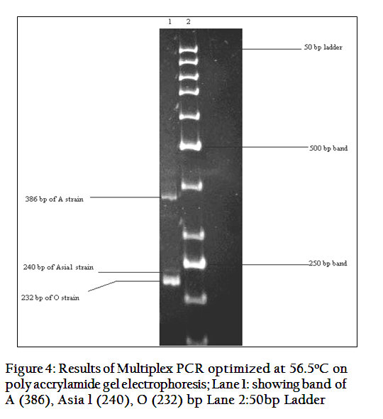
Figure 4: Results of Multiplex PCR optimized at 56.5oC on poly accrylamide gel electrophoresis; Lane 1: showing band of a (386), Asia l (240), O (232) bp Lane 2:50bp Ladder
DISCUSSION
In this study we found mPCR a very quick and reliable method to identify the specific serotype of FMD in any filed outbreak. A 328 bp fragment was amplified for all three serotypes (A, O and Asia1) using universal primer pair (IF: IR). Similarly a study was carried out by Reid et al (2000) using same primers (1F:1R); their results had also an amplicon of 328 bp. Alamdari et al. (2006) used phenol–chloroform thiocyanate–based method for RNA extraction rather than trizole reagent. Giridharan et al., (2005) isolated RNA using kit (Qiagen, Germany). The difference in RNA extraction protocol has been found to be no effect on the results. For cDNA synthesis instead of random hexamers they used oligonucleotide primers. Their results were different giving a band size of 131 bp. Difference in results was due to the use of different primer pair for genome detection of FMDV. For serotype identification and primers optimization of A, O and Asia1, PCR was carried out using serotype specific primers designed against VP1 gene in individual reaction mixture of 25 μl (A1F:A1R, O1F:O1R, AS1F:AS1R). Amplicon size of 386, 232 and 240 bp for A, O and Asia1 serotypes were obtained respectively in current study. Giridharan et al. (2005) designed primers against VP1 gene native to their country (serotype specific) and capsid–coding region of FMDV. Their results for O, A, Asia1 were 249, 376 and 537 bp respectively. Difference was due to the primers’ positions giving different amplicons as compared to the present study.
Then optimization of PCR cycling conditions for A, O and Asia1 serotypes of FMDV done individually. Among the cyclic conditions, increase of annealing temperature from 56 to 60°C did not alter the amplification efficiency but above 60°C, it was affected adversely giving non specific products. At primer to template concentration/ratio of 0.5μl, 1μl and 1.5μl, to 1.5μl, 2μl and 3μl template, product was amplified at primer concentration of 1.5μl (forward and reverse) and template concentration of 3.0μl whereas; in other combinations non specific products were obtained. The number of cycles were only increased from 30–40 for that template DNA which has higher GC contents.
Callens and De Clercq (1997) used primers for the amplification of A, O and Asia1 serotypes of FMDV. Primers’ sequences were different from the present study. They designed from the 1D and 2AB genes of 1C region of the FMD viral genome. Primers amplified the fragments 402, 732and 296 bp size for O, A and Asia 1 serotypes. As success of any PCR is dependent on the efficiency of primer and template binding, it would be ideal if the primers were designed on the virus sequences native to that particular geographical area.
Multiplex PCR was optimized to amplify the three major serotypes of FMDV A, O and Asia 1 in a single reaction. Primers were designed against the VP1 gene of virus. Initially the primers were optimized individually for each serotype by using gradient temperature PCR. Then all three serotypes were subjected to single multiplex PCR reaction. Annealing temperature for this reaction was optimized at 56.5°C. The amplicon sizes obtained were 386 bp, 240 bp and 232 bp for A, Asia 1 and O serotypes respectively. In mPCR primer to template ratio was optimized. The primer concentration was kept constant at 1.5 μl of 10mM of each forward and reverse primer total 9 μl for three serotypes whereas; template DNA ratio was changed. At template concentration of 1μl, 1.5μl and 2μl non specific products were obtained. The non specific products were obtained because of less quantity of template DNA resulting in primer dimerization. The use of 3μl of cDNA for each serotype and at annealing temperature of 56.5°C has given specific PCR products. At temperatures 53–60°C, specific amplification of the target sequence was found in all the serotypes without much variation in band intensity. The annealing temperature in the study of Alamdari et al., (2006) was optimized at 600C of mPCR. The specific primers were selected from the 2B region of FMDV genome. DNA Fragments of 292, 402 and 732 bp were amplified for the Asia1, O and A serotypes, respectively. There was no difference between amplicon size of serotype O and Asia 1 of FMDV, because of 8 kbp difference between two serotypes that were resolved on 1.5% agarose gel electrophoresis. To resolve this issue amplicon of mPCR were analyzed on 3% PAGE.
The sensitivity of mPCR was determined by making 10–fold serial dilutions of extracted RNA. The serotype A and Asia1 showed the results upto 103 dilution with decreasing band intensity whereas; with serotype O results obtained up to 104 dilution. This is because sensitivity depends upon length of primer and template DNA concentration. Primers with shorter length were comparatively sensitive than lengthy primers. The specificity of the test in differentiating FMDV infection from other vesicular infections could be validated experimentally. The test specifically detects FMDV infection because primers were designed only against the FMDV genome. The field samples in mPCR, produced a single band of Asia1 serotype of FMDV. Woodbury et al. (1994) analyzed field specimen (Sau 8/88) from Saudi Arabia and reported that O serotype was dominant followed by Asia 1 to some extent. The results were different from present study due to varied geographical distribution of different serotypes.
It is evident from the present study that multiplex PCR is a quick, time saving, cost effective and efficient method to detect different subtypes in a single reaction. It is therefore a helpful tool for the diagnosis of FMDV leading to its effective control and prevention.
REFERENCES
Alamdari MS, Ghorashi, Salehi–Tabar MAR (2006). Detection of foot–and–mouth disease virus and identification of serotypes in East Azerbaijan province of Iran. Veternarski Archiv. (5): 413 – 419.
Anjum R, Hussain M, Zahoor AB, Irshad H, Farooq U (2006). Epidemiological analyses of Foot and Mouth disease in Pakistan. Int. J. Agr. Biol. 5: 648 – 651.
Callens M, Clercq DK (1997). Differentiation of the seven serotypes of foot–and–mouth disease virus by reverse transcriptase polymerase chain reaction J. Virol. Methods. (67): 35 – 44.
http://dx.doi.org/10.1016/S0166-0934(97)00074-8
Chomczynski P, Sacchi N (1987). Single step method of RNA isolation by acid guanidinium thiocyanate–phenol–chloroform extraction. Ann. Biochem. 162(1): 156 – 159.
http://dx.doi.org/10.1006/abio.1987.9999
http://dx.doi.org/10.1016/0003-2697(87)90021-2
Giridharan P, Hemadri D, Tosh C, Sanyal A, Bandyopadhyay SK (2005). Development and evaluation of a multiplex PCR for differentiation of foot–and–mouth disease virus serotypes native to India. J. Virol. Methods. 126 (1–2): 1 – 11.
http://dx.doi.org/10.1016/j.jviromet.2005.01.015
PMid:15847913
Hall MD, Knowles NJ, Wadsworth J, Rambaut A, Woolhouse MEJ (2013). Reconstructing geographical movements and host species transitions of foot–and–mouth disease virus serotype SAT 2. mBio 4(5): e00591–13
Hindson BJ, Scott MR, Brian RBr, Katja E, Nigel PF, Lance FBT, Raymond JL, Pejman N, Elizabeth AV, Thomas RS, Pamela JH, Donald PK (2008). Diagnostic Evaluation of Multiplexed Reverse Transcription–PCR. Microsphere Array Assay for Detection of Foot–and–Mouth and Look–Alike Disease Viruses. J. Clin. Microbiol. 46(3): 1081 – 1089.
http://dx.doi.org/10.1128/JCM.01740-07
PMid:18216216 PMCid:PMC2268367
Knowles NJ, Samuel AR (2003). Molecular epidemiology of foot–and mouth disease virus. Virus Res. 91: 65 – 80.
http://dx.doi.org/10.1016/S0168-1702(02)00260-5
Morioka K, Fukai K, Yoshida K, Yamazoe R, Onozato H, Ohashi S, Tsuda T, Sakamoto K (2009). Foot–and–Mouth disease virus antigen detection Enzyme–Linked Immunosorbent Assay using multiserotype– reactive monoclonal antibodies. J. Clin. Microbiol. 47(11): 3663 – 3668.
http://dx.doi.org/10.1128/JCM.00695-09
PMid:19759230 PMCid:PMC2772642
OIE (2012). OIE, Foot and Mouth Disease In: Manual of Diagnostic Test and Vaccines for Terrestrial Animals, Chapter, 2.5.
Reid SM, Ferris NP, Hutchings GH, Samuel AR, Knowles NJ (2000). Primary diagnosis of foot–and–mouth disease by reverse transcription polymerase chain reaction. J. Virol. Methods. (89): 167 – 176.
http://dx.doi.org/10.1016/S0166-0934(00)00213-5
Sangula AK, Siegismund HR, Belsham GJ, Balinda SN, Masembe C, Muwanika VB (2011). Low diversity of foot–and–mouth disease serotype C virus in Kenya: evidence for probable vaccine strain re–introductions in the field. Epidemiol. Inf. 139: 189 – 196.
http://dx.doi.org/10.1017/S0950268810000580
PMid:20334728
Tosh C, Sanyal A, Hemadri D, Venkataramanan R (2002). Phylogenetic analysis of serotype A foot–and–mouth disease virus isolated in India between 1977 and 2000. Arch. Virol. 147: 493 – 513.
http://dx.doi.org/10.1007/s007050200002
PMid:11958451
Wiesław N, Kęsy A (2010). Rapid detection and quantification of foot–and–mouth disease virus by a real–time reverse transcription PCR. Bull. Vet. Inst. Pulawy, 54: 3–7.
Woodbury EL, Samuel AR, Knowles NJ, Hafez SM, Kitching RP (1994). Analysis of mixed foot–and–mouth disease virus infections in Saudi Arabia prolonged circulation of an exotic serotype. Epidemiol. Inf. 112 (1): 201 – 211.
http://dx.doi.org/10.1017/S0950268800057575
PMid:8119359 PMCid:PMC2271471
Xu L, Hurtle W, Rowland JM, Casteran KA, Bucko SM, Fred R, Valdazo–González GB, Knowles NJ, King DP, Beckham TR, McIntosh MT (2013). Development of a universal RT–PCR for amplifying and sequencing the leader and capsid–coding region of foot–and–mouth disease virus. J. Virol. Methods 189: 70 – 76.
http://dx.doi.org/10.1016/j.jviromet.2013.01.009
PMid:23380590


