Advances in Pharmaceutical and Ethnomedicines
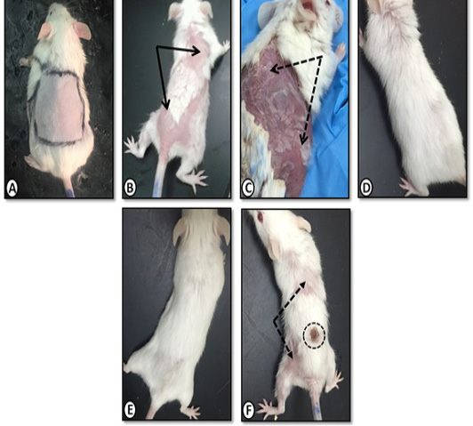
Gross morphology of mice dorsal back skin of A. Control group with normal skin appearance. B. Benzene painted (two weeks) showing patchy hair loss (arrows)and redness (erythema). C. Benzene painted (three weeks) showing skin dryness and scaling (dotted arrows). D. Achillea in olive oil treated skin showing normal skin with no dryness or scales E. Olive oil treated skin with similar improvement. F. Medicinal cream (Metazone) treated showing moderate improvement, still scaling (dotted arrow) and hair loss. Dotted circle (site of skin biopsy).
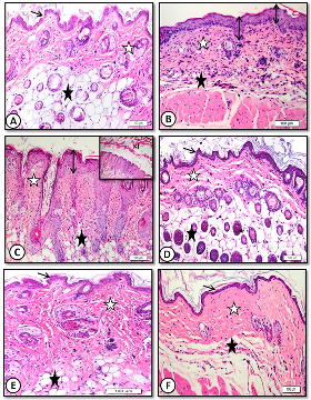
Low magnification (x20) of mice dorsal back skin stained with H&E showing A. control with normal epidermal thickness (thin arrows), dermis (white star) and hypodermis (black star). B .benzene painted (two weeks) increased epidermal thickness (arrows) and dermal inflammatory cells with decreased adipose tissue (black star). C. benzene painted (two weeks) marked increase in epidermal thickness and keratin layer (KL) with desquamation. Note ridge elongation (double head arrows). D. Achillea in oil treated skin showing normal epidermal thickness (arrows) and dermis (white star) with normal hair follicles and sebaceous glands (black stars). E. Oliveoil treated skin with similar improvement. F. medicinal cream treated skin with normal epidermal thickness(arrow), few hair follicles and inflammatory cells (stars).

High magnification (x60) of mice dorsal back skin stained with H&E showing: A. control with normal epidermal thickness (double heads arrow). B. Benzene painted skin (three weeks) showing increase in epidermal thickness (double head arrow),dermis (white star) is infiltrated with mononuclear and mast cells (dotted arrows). C. Benzene painted skin (three weeks) showing marked increase in epidermal thickness and ridge elongation (double head arrows). D. Achillea in oil treated showing normal epidermal thickness, hair follicles and sebaceous glands (white star) with absence of inflammatory cells (black star). E. Olive oil treated skin showing normal epidermal thickness (double head arrows), few sebaceous glands (white star) and inflammatory cells (black star). F. Medicinal cream treated skin with normal epidermal thickness (double head arrow), few hair follicles and inflammatory cells (stars).
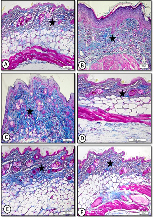
Low magnification (x20) of mice dorsal back skin stained by Masson trichrome for collagen fibers showing A. Control with normal collagen content (star). B. Benzene painted with increase collagen(star). C. Benzene painted (three weeks) with increase collagen content (star). D. Achillea in oil treated showing normal collagen content (star). E. Olive oil treated skin showing normal collagen content (star). F. Medicinal cream treated skin with normal collagen content (star).
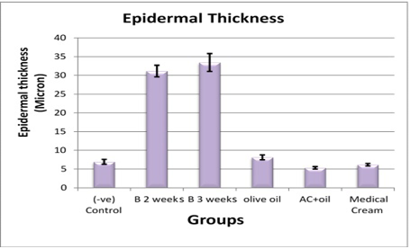
Epidermal thickness in control and all treated groups. AC, Achillea, B, benzene.


