Journal of Animal Health and Production
Case Report

Non-Cerebral Coenurosis in Goats-A Case Study
Venkatesan Margandan1, Saravanan Mani2, Selvaraj Palanisamy1, Yogeshpriya Somu1, Jayalakshmi Konappan1, Veeraselvam Muthusamy1
1Department of Veterinary Medicine, Veterinary College and Research Institute, Orathanadu – 614 625, Tamil Nadu Veterinary and Animal Sciences University (TANUVAS); 2Teaching Veterinary Clinical Complex, Veterinary College and Research Institute, Orathanadu – 614 625, Tamil Nadu Veterinary and Animal Sciences University (TANUVAS), India.
Abstract | A rural goat farm with 93non-descript goats were investigated for the complaint of emaciation, weight loss and fluctuating palpable masses in various parts of the body. Out of 93 goats, 12 were found to have cysts on dorsal lumbar, right flank and prescapular areas and identified as Coenurus gaigeri. All the goats were treated with Fenbendazole 7.5mg/kg orally.
Keywords | Non-cerebral coenurosis, Goats, Coenurus gaigeri
Editor | Asghar Ali Kamboh, Sindh Agriculture University, Tandojam, Pakistan.
Received | March 05, 2018; Accepted | March 25, 2018; Published | March 30, 2018
*Correspondence | Venkatesan Margandan, Department of Veterinary Medicine, Veterinary College and Research Institute, Orathanadu – 614 625, Tamil Nadu Veterinary and Animal Sciences University (TANUVAS), India; Email: drvenksmvsc88@gmail.com
Citation | Venkatesan M, Saravanan M, Selvaraj P, Yogeshpriya S, Jayalakshmi K, Veeraselvam M (2018). Non-cerebral coenurosis in goats-a case study. J. Anim. Health Prod. 6(1): 47-50.
DOI | http://dx.doi.org/10.17582/journal.jahp/2018/6.1.47.50
ISSN | 2308-2801
Copyright © 2018 Venkatesan et al. This is an open access article distributed under the Creative Commons Attribution License, which permits unrestricted use, distribution, and reproduction in any medium, provided the original work is properly cited.
INTRODUCTION
Coenurosis is a serious disease responsible for high economic losses in sheep and goat farm, in addition to its zoonotic impact. C. cerebralis considered as the principal cause for nervous manifestations in sheep and goats (Desouky et al., 2011). The parasite responsible for non-cerebral coenurosis was initially named Multiceps gaigeri (Hall, 1916) in goats and M. Skrjabini in sheep (Schuster et al., 2010) and later it was considered M. gaigeri as the same species with T. multiceps (Verster, 1969; Soulsby, 1982).This study reports the non cerebral form of coenurosis (dorsal lumbar, right flank and prescapular areas) and its clinical management in goats.
MATERIALS AND METHODS
A referral clinical investigation was under taken in arural goat farm with 93 non-descript goats during November 2017 in Thanjavur, Tamil Nadu. The farm was reported to have complaints of weight loss in animals and palpable fluctuating masses in various parts of body. No deworming and vaccination were done by the farmer. On clinical examination, vital signs were normal except for pale conjuctival mucous membrane in all the animals. Physical examination revealed subcutaneous fluctuating palpable masses of various sizes on the dorsal lumbar, right flank and prescapular areas (Figure 1a, b & c). Other systemic examinations were found to be apparently normal. During the early phase, one goat was presented to the Large Animal Medicine Referral Clinic and an ultra-sonographic assessment of the mass was done. It was found to be a cyst and ultrasound guided aspiration of it revealed presence of coenurus. Suspecting the spread of coenurosis, aspiration, incisions were made over the masses and samples were collected. Microscopic and macroscopic examination of the aspirate and brood capsules revealed the presence of Coenurus gaigeri.
RESULTS AND DISCUSSION
Out of 93 goats examined for the presence of cysts on the body, only 12 goats had cysts on dorsal lumbar, right flank and prescapular areas (Figure 1a, b & c) and it was identified as Coenurus gaigeri, which is an intermediate stage of T. Multiceps gaigeri.
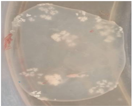
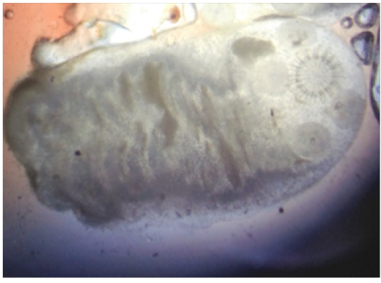
Goat with history of restricted gait, inappetence, respiratory distress and retarded growth found to have more than 200 C. gaigeri because of that animal was “puffed up” in appearance reported by Dey et al. (1988). In non-cerebral coenurosis of goats there will be lameness, paresis and paralysis together with outgrowing skin lumps, due to the subcutaneous cysts, are the major clinical manifestation (Ramadan et al., 1973; Oryan et al., 2010, 2012). In the present study, the animals despite having their parasitism, would appear healthy. The only clinical abnormality observed in infected goats was the palpation of variable sizes of single cyst in dorsal lumbar, right flank, prescapular regions. In cerebral coenurosis interpretation of the clinical signs in combination with detailed location of the cyst, using ultrasound examination is the best method of diagnosis (Skerritt and Stallbaumer, 1984; Tirgari et al., 1987; Biswas, 2013). Subcutaneous/ intramuscular presence of cyst in one goat, which presented to the Large Animal Medicine Referral Clinic was confirmed ultra-sonographically and ultrasound guided aspiration of it revealed presence of coenurus. Aspiration and incised masses having brood capsule contained thick, transparent fluid, with number of small size white colour plaques (Figure 2). Microscopic view of single scolex showed typical taenia hooks (Figure 3).
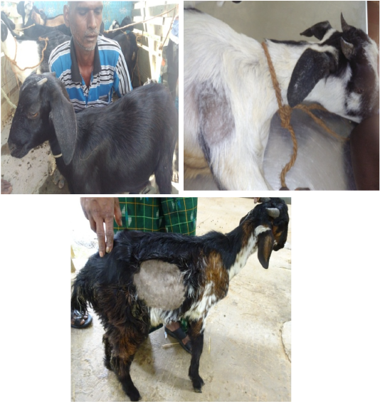
Figure 1: a) Cyst on the dorsal lumbar region; b) Cyst in the prescapular region; c) Cyst in the right flank region behind the last rib
The larval stage coenurus (C. cerebralis) commonly affects the central nervous system (CNS), particularly the brain and gives rise to the neurological signs (Soulsby 1982). In addition to infecting the brain, the larval stage also develops in the subcutaneous tissues, muscles and some times in the body cavities (Schuster et al., 2010). Various authors were reported presence of cysts in different locations of goat’s body; a cyst was found over the right mandible (Devasana et al., 1998), muscles of the thighs (Jyothimol and Sangaran, 2015), hips and shoulders (Ramadan et al., 1973), neck region, in connective tissue near the splenic ligament and in intercostals and abdominal musculature (Shastri et al., 1985). It was also reported in visceral cavity including heart, diaphragm, thoracic cavity, abdominal cavity and pelvic inlet, pericardium and myocardium (ICAR, 2016-17), and in intramuscular and subcutaneous tissues of goats (Oryan et al., 2014). Shivapraksh and Reddy (2009) also found multiple subcutaneous coenuri in neck, prescapular region, abdomen and limbs in a herd of goats as C.gaigeri because of their extra-cranial locations. In the present study, the coenurus cysts were fluctuating andfoundsubcutaneously of various sizes on the dorsal lumbar, right flank and prescapular region. This may reflect a different host response to the parasite in goats or, alternatively, parasitism by larvae of another cestode species, T. Gaigeri (Verster 1969; Sharma et al., 1995; Moghaddar, 2007). The cerebral form of the cysts produced by T. multiceps in sheep is genetically identical with the non-cerebral cysts of T. multiceps gaigeri (Verster 1969; Sharma et al., 1995) in intramuscular and/or subcutaneous tissues in goats (Moghaddar, 2007; Christodoulopoulos et al., 2015).
Albendazole or combinations of anthelmintics (Fenbendazole and Praziquantel) were useful in coenurosis (Ghazaei, 2005). In the current study, the animals were treated with suspension of Fenbendazole and Praziquantel @ 7.5mg per kg body weight orally and herbal liver tonics were given to increase the immunity as well as appetite. Review of the farm a week later confirmed that the animals were doing well. Albendazole or combinations of anthelmintics (Fenbendazole and Praziquantel) were useful in coenurosis (Ghazaei, 2005).
Lack of community dogs management and stray dog control programme in villages, coupled with lack of awareness and knowledge about the impact of coenurosis and the free access for dogs to eat offals at village meat selling points enables easy spread of coenurosis. Absence of village level community animal health programs to take care of periodical deworming and vaccinations also facilitate occurrence of coenurosis. All of which are further enhanced by the fact that there were no deworming of village dogs as well as stray dogs of the locality. This further contributes to the persistent spread of coenurosis.
CONCLUSION
Aspiration/incision techniques and ultra-sonographic guided aspirations helped in diagnostic planning and for further treatment of non-cerebral coenurosis in goats. Current case study highlighted that the lack of periodic basic health care practices like deworming in dogs resulted in an easily preventable disease (coenurosis) besides developing into a complicated entity and posing challenges for the practicing veterinarians and economic losses to the poor people.
Acknowledgments
The authors acknowledge the support of The Dean, Veterinary College and Research Institute, Orathanadu, Tamil Nadu, India to carry out the study.
Conflict of interest
There is no conflict of interest
Authors Contribution
Venkatesan Margandan and Saravanan Mani: Disease investigation, Data collection, literature and preparation of manuscript.
Selvaraj Palanisamy: Guided the case study and Final proof reading of the manuscript.
Yogeshpriya Somu and Jayalakshmi Konappan: Sample (Cyst) processing and identification of Coenurus gaigeri.
Veeraselvam Muthusamy: Preparation of manuscript and assisting the treatment.
References


