Journal of Animal Health and Production
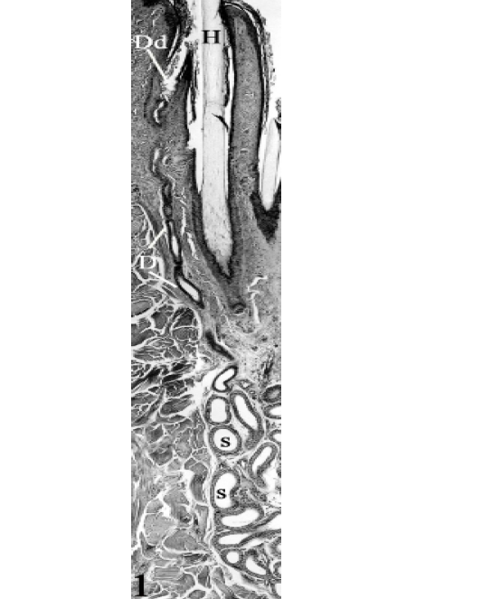
An epitrichial sweat gland at the thigh region of seven years-old camel , consisting of two portions: the secretory portion (S) and the excretory duct (D) opening into the primary hair follicle in the nearby of the epidermis. Dilated destination device (Dd), primary hair (H). H&E, X100.
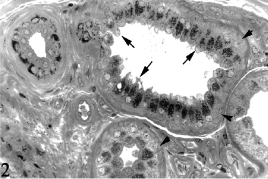
The secretory end-pieces of the epitrichial sweat glands at the thigh region of 6 years-old camel, showing numerous basophilic granules at the apical portion of the cells. Some cells bear a budding appearance (Cytoplasmic blebbing, arrows) apically. Myoepithelial cells (Arrowheads). Toluidine blue, X400.

3A, The secretory portion of the epitrichial sweat gland at the belly region of 6 months-old camel, showing the commencement of the excretory duct (D), secretory portion (S). Toluidine blue, X160. Inset: The excretory duct at the thigh region of 6 years-old camel, showing a wavy pattern in their way to open into a primary hair follicle. Toluidine blue, X250. 3B, The transition between the secretory portion (S) and the excretory duct (D) at the neck region, of 3 years-old camel. Toluidine blue X160. 3C, The excretory duct of the epitrichial sweat gland at the shoulder region, towards its opening into the primary hair follicle (upwards), Ap= Arrector pili muscle. Toluidine blue, X160. 3D, The excretory duct (arrow), receiving a conical jacket of stratified epithelium, in its way to open into the apex of the destination device, sebaceous gland (Sg). Toluidine blue, X 250.
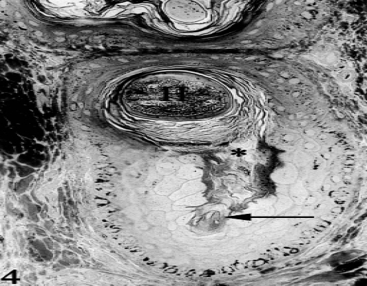
Cross section of the hair follicle at the level of the dilated destination device, at the thigh region, of six years-old camel, showing the nozzle-shaped terminal portion of the excretory duct (arrow) and the opening of this device into a neighbouring hair follicle (*). Notice the histomorphological differences between the epithelial lining of the hair follicle and that of the dilated device. Primary hair (H), Toluidine blue, X250.
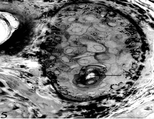
Tangential section of the destination dilated device, showing the peculiar cellular elements of the lining epithelium, at the thigh region of six years-old camel. Excretory duct (arrow), Toluidine blue, X250.
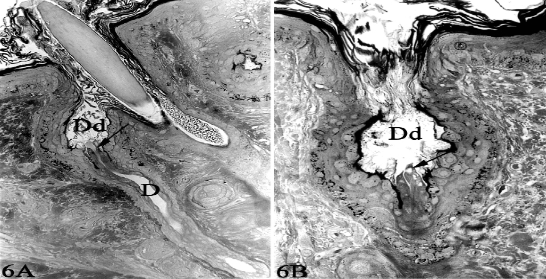
The terminal portion of the excretory duct (D) opening into an oval or rounded-shaped dilated destination device (Dd) at the outer most portion of a primary hair follicle (epitrichial sweat gland “A”) or emptying on to the skin surface in an interfollicular location (atrichial sweat gland “B”). Notice the terminal portion of excretory duct (arrow) projecting into the lumen of the dilated destination device. Toluidine blue, X250.


