Journal of Animal Health and Production
Research Article

Seroprevalence and Histopathological Study of Neosporosis in Water Buffaloes (Bubalus bubalis) in Baghdad City, Iraq
Amer Murhum Al-Amery1*, Azhar Ali Faraj1, Inam Bader Faleh2
1Department of Parasitology; 2Department of Pathology, Faculty of Veterinary Medicine, University of Baghdad, Iraq.
Abstract | The current study was designed to identify the seroprevalence of Neospora caninum as well as the effect of age, sex and months on the infection rate in water buffaloes (Bubalus bubalis) in Baghdad city. One hundred and fifty blood samples were collected during October 2014 to May 2015. Results showed an overall infection rate of 20% analyzed by enzyme linked immunosorbent assay. The age showed a significant association (P≤0.05) with the prevalence of N. caninum infection. The highest infection rate (32%) was shown in the age group <3-5 years old, whereas no infection was reported in the age group <1-2 year old. There was no significant effect (P≥0.05) of sex factor on the infection rate of N. caninum. The histological analysis revealed significant lesions in different internal organs i.e., brain, heart and lung of aborted fetuses and placentome of naturally infected dam. The main lesions observed in these tissue were congested blood vessels, presence of pseudocysts containing number of bradyzoites and intra muscular edema. This is the first report on neospora antibodies in water buffaloes from Baghdad city that could be correlate with abortions and neonatal mortality in water buffaloes of study area, causing devastating economic losses to the beef and dairy industries.
Keywords | Seroprevalence, Neoospora caninum, Buffaloes, Abortion, Neosporosis, Histopathological study
Editor | Asghar Ali Kamboh, Sindh Agriculture University, Tandojam, Pakistan.
Received | June 10, 2016; Accepted | July 10, 2016; Published | July 28, 2016
*Correspondence | Amer M Al-Amery, Department of Parasitology, Faculty of Veterinary Medicine, University of Baghdad, Iraq; Email: m.murhum@yahoo.com
Citation | Al-Amery AM, Faraj AA, Faleh IB (2016). Seroprevalence and histopathological study of neosporosis in water buffaloes (Bubalus bubalis) in Baghdad city, Iraq. J. Anim. Health Prod. 4(3): 101-104.
DOI | Http://dx.doi.org/10.14737/journal.jahp/2016/4.3.101.104
ISSN | 2308–2801
Copyright © 2016 Al-Amery et al. This is an open access article distributed under the Creative Commons Attribution License, which permits unrestricted use, distribution, and reproduction in any medium, provided the original work is properly cited.
Introduction
Neospora caninum is an obligate protozoan parasite which causes abortion in farm animals worldwide especially in cattle. The parasite was first detected in 1984 in dogs (Dudey et al., 2007; Dubey and Schares, 2011). Domestic and wild canids are natural final host for N. caninum. Infected dog excrete oocysts in their feces, which is then ingested by intermediate hosts such as horses, goats, sheep and water buffaloes (Nasir et al., 2011; Kamali et al., 2014).
The disease is generally diagnosed in bovine by serological or pathological findings, immunochemical methods and molecular biological techniques. To detect antibodies specific for N. caninum tachyzoites, antigens are examined using serological test, such as enzyme linked immunosorbent assay (ELISA) and indirect fluorescent antibodies test (IFT) (Bishop et al., 2010; Rhman et al,. 2011; Jamal and Haidar, 2014). In Iraq less is known about seroprevalence of neosporosis in bovine. The purpose of this study was to identify the prevalence of N. caninum antibodies in sera of water buffaloes in Baghdad city.
Materials and Methods
Blood Collection and Analysis
One hundred and fifty blood samples were collected randomly from different ages and sexes (60 males and 90 females) of water buffaloes during October 2014- May 2015 from different areas north of in Baghdad city, Iraq. The samples were transported on cold box to the parasitology laboratory, College of Veterinary Medicine, Baghdad University. Blood samples (about 5 ml) were collected by puncturing external jugular vein under a septic condition. Blood samples were transferred to tubes without anticoagulant additive and centrifuged at 1000 rpm for 15 min, then serum was stored at -20°C until analyzed. The blood samples were examined by ELISA according to manufacturer instruction (IDvet, France).
Histopathological Examination
Biopsy from brain, heart and lung were collected from 5 day old aborted fetus and placenta by seropositive doe from infected dam, fixed in 10% buffered formalin solution for (2-3) days, then routinely processed in histokinette. Tissue section were embedded in paraffin, and sections of 5 µm thickness were cut by microtome and stained with hematoxylin and eosin then examined under light microscope (Luna, 1968).
Statistical Analysis
Data was statistically analyzed by Chi-square tests for significance using SPSS 15 version.
Table 1: Seroprevalence of neosporosis infection in water buffaloes analyzed by ELISA
|
Sample |
No. of samples examined |
Infected |
|
|
Number |
Percentage |
||
|
Serum |
150 |
30 |
20 |
Table 2: Seroprevalence of neosporosis infection in water buffaloes in different months
|
Months |
No. of samples examined |
Seropositive |
|
|
Number |
Percentage |
||
|
October2014 |
18 |
4 |
22.22 |
|
November |
20 |
4 |
20 |
|
December |
19 |
4 |
21.05 |
|
January2015 |
18 |
3 |
16.66 |
|
February |
19 |
4 |
21.05 |
|
March |
20 |
4 |
20 |
|
April |
16 |
3 |
18. 75 |
|
May |
20 |
4 |
20 |
P≥0.05
Results and Discussion
Table 1 shows an overall infection rate in serum of water buffaloes as 20%, while other researchers reported that the seropositivity in water buffaloes was 67% in Iran (Mohammed et al., 2007), 4.5% in Northeastern Thailand (Nam et al., 2012), 42.8% in Pakistan (Naser et al., 2014) and 47% - 59% in Southern Italy (Borriello et al., 2014). This variation in the infection rate between the countries may be due to difference in the numbers of examined animals, survey periods and serological testes used. Moreover, climatic factors also affect the abundance of viable parasitic stages in the environment for final and intermediate hosts (Ahmet et al., 2014).
Furthermore, study results showed no statistical differences (P≥0.05) in total infection rates of N. caninum during the months of the study (Table 2). This could be due to that the seasonal prevalence is difficult because antibodies against N. caninum can persist for several months (Jung et al., 2014; Al-Saadi, 2015).
A high rate of infection (32%) appeared in the age <3-5 years old, while the lowest prevalence (14.2%) was observed in <2-3 years old buffaloes. Also no infection rate was recorded at the age group <1-2 years old (Table 3). These results were in accordance with those found in previous studies which showed that the prevalence appeared at the age group of above 4 years old (Mallah et al., 2012; Al-Jomaily and Al-Rubaie, 2013). The high rate of infection among these age groups were may be due to ingestion of oocysts that shed by definitive host. In addition to that, the results of present study showed no infection rate in the age group <1-2 years old, that may be due to maternal immunity or less exposure to the parasite oocysts (Al-Jomaily and Al-Rubaie, 2013).
Table 3: Seroprevalence of neosporosis infection in different age groups of water buffaloes
|
Age |
No. of samples examined |
Seropositive |
|
|
Number |
Percentage |
||
|
<1-2 |
25 |
0 |
0 |
|
<2-3 |
35 |
5 |
14.2* |
|
<3-5 |
50 |
16 |
32* |
|
<5-6 |
40 |
9 |
22.5* |
*Values differ significantly (P≤0.05)
Current study also shows a higher infection rate in female (22.22%) than in males (16.66%) without significant difference (Table 4), that agreement with Hamid and Gorani (2007) who demonstrated no significant differences between sexes. This could be due to both sexes exposed to the same environmental condition.
Table 4: Seroprevalence of neosporosis infection in water buffaloes according to gender
|
Gender |
No. of samples examined |
Seropositive |
|
|
Number |
Percentage |
||
|
Male |
60 |
10 |
16.66 |
|
Female |
90 |
20 |
22.22 |
P≥0.05
The main characteristic lesions of N. caninum infection in the placenta represented was polymorphonuclear cells infiltration and blood vessels congestion (Figure 1A) and presence of pseudocyst containing several bradyzoites (Figure 1B). The lesion in the brain showed large necrotic cavities containing tissue debris indicating areas of encephalomalacia seen with number of degenerating astrocysts (Figure 2).
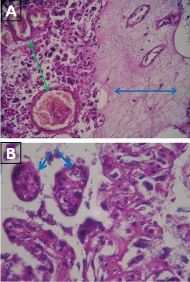
Figure 1: Histopathological sections in placentome of infected dam: A) Fibrinous precipitation (green arrow), polymorphonuclear cells infiltration and blood vessels congestion (blue arrow) (H&E stain x 40); B) Presence of pseudocyst containing several bradyzoites (blue arrow) (H&E stain x40)
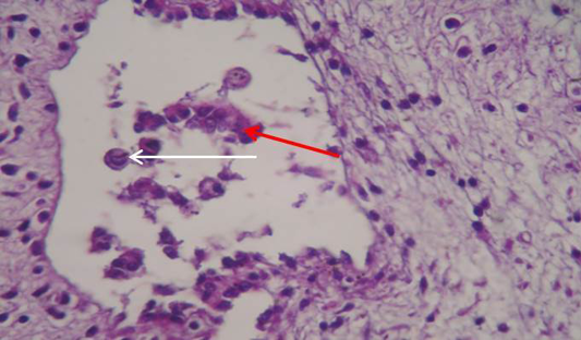
Figure 2: Showing section of brain of aborted fetus. Large necrotic cavities containing tissue debris indicating areas of encephalomalacia (Red arrow) with number of degenerating astrocysts (White arrow) (H&E stain x40)
It was demonstrated massive fragmentation and separation of myocardial bundles with intra muscular edema resulting in severe atrophy of muscle bundles in the heart (Figure 3).The pathological lesion in the lung showed severe blood vessels congestion associated with interstitial hemorrhage with presence of pseudocysts containing several bradyzoites (Figure 4). The evaluation of pathological lesions in the placentome of infected dam and brain, heart and lung of aborted fetus expressed high level of anti-Neospora caninum antibodies. This observation may indicate that the animals were exposed to parasite N. caninum, as suggested previously (Auriemma et al., 2014; Mallah et al., 2012). These studies also reported necrosis in many organs during the presence of parasite in organs after the inflammatory reaction against the parasite N. caninum which is capable of producing grossly visible lesion. Generally, the pathologic features of systemic neosporosis include pneumonia, nervous system disease and myocarditis may occur (Dubey and Lindsay, 1990; Dubey, 2003).
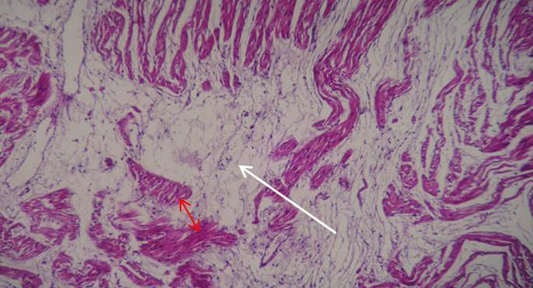
Figure 3: Showing section of heart of aborted fetus. Massive fragmentation and separation of myocardial bundles with intra muscular edema (White arrow) resulting in severe atrophy of survival muscle bundles (Red arrow) (H&E stain x40)
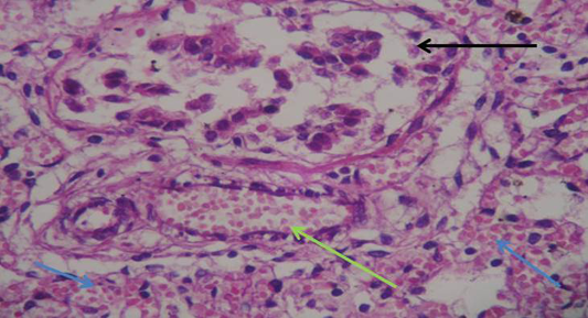
Figure 4: Showing section of lung of aborted fetus. Severe bloods vessels congestion (green arrow) associated with interstitial hemorrhage (blue arrow) with presence of pseudocysts containing several bradyzoites (black arrow) (H&E stain x40)
Conclusion
Current study reported the first time detection of Neospora caninum antibodies in serum of water buffaloes in Baghdad-Iraq. The role of Neospora caninum as a causative agent of abortions in animal species of study area could be established. Consequently, neosporosis screening should be the routine diagnosis of abortive agents in water buffalo, especially in herds characterized by pregnancy interruption or fertility loss.
Acknowledgments
My thanks to all staff of Parasitology and Pathology Departments in College of Veterinary Medicine, University of Baghdad, Iraq.
Conflict of interest
The author declares that there is no conflict of interest.
Authors’ Contribution
Amer Murhum and Azhar conducted sample collocation and serological test. Inam Bader conducted histobathological test.
REFERENCES


