Journal of Animal Health and Production
Research Article
J. Anim. Health Prod. 9(4): 479-486
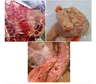
Figure 1
Bovine tuberculosis lesions discovered in slaughterhouses. Pleura with solid spherical formations covered with yellowish nodules (Pearl disease) (a), tracheobronchial Ln section with yellowish caseous consistence (b), digestive tract showing TB lesion in the rumen wall(c).
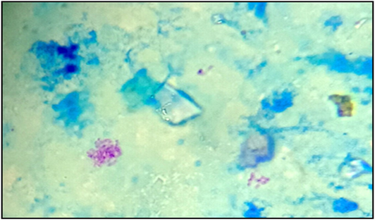
Figure 2
Acid-fast bacilli appeared as pinkish coccobacilli in a bluish background detected in lymph nodes from calve (x100).
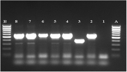
Figure 3
Agarose gel electrophoresis of the amplified MTC product. A, B: DNA marker (80-1000pb), 1: negative, control, 2: positive control M.bovis (BCG), 3: positive control M.tuberculosis (H37Rv), 4-8: MTC PCR product.


