Journal of Animal Health and Production
Research Article

Serodiagnosis of Foot and Mouth Disease Antibodies in Sheep and Goat Sera by using NSP-cELISA in Garmian Region, Kurdistan, Iraq
Osman M. Jabary*, Hiwa Yassin Essa, Yousef Khalel Bawakhan, Salim Sabah Abdullah, Nisreen Hadi Ali, Mohammed Tharwat Kareem, Shilan Abubakir Mohammed Amin
Directorate of Veterinary in Garmian, Iraq.
Abstract | A serosurvey was carried out by using cELISA (competitive enzyme linked immunosorbent assay) to detect the presence of foot and mouth disease (FMD) antibodies in apparently healthy small ruminants (sheep and goats) in Garmian region, Kurdistan, Iraq. The test is valuable in serological surveys because only infected animals with FMD virus will give positive reaction. A total 184 of serum samples (142 sheep and 42 goats) were collected randomly from eighteen flocks belongs to fourteen villages. The result revealed that 36.95% of animals (39.43% sheep and 28.57% goats) show positive results to ELISA antibodies. Moreover, the results were also indicated that 36.77% of female and 37.93% of male animals appeared positive. Furthermore, the high seropositive rate was detected at age >3 years (44 %), that followed by 40% and 14.28% at 1-3 years and <1 year age, respectively. In conclusion, the significant proportion of small ruminants of Garmian region of Iraq is seropositive for FMD.
Keywords | FMD, cELISA, Seroprevelence, Small ruminant, Garmian, Kurdistan, Iraq
Editor | Asghar Ali Kamboh, Sindh Agriculture University, Tandojam, Pakistan.
Received | December 16, 2019; Accepted | March 30, 2020; Published | May 04, 2020
*Correspondence | Osman M. Jabary, Directorate of Veterinary in Garmian, Iraq; Email: dr.osmanmh@yahoo.com
Citation | Jabary OM, Essa HY, Bawakhan YK, Abdullah SS, Ali NH, Kareem MT, Amin SAM (2020). Serodiagnosis of foot and mouth disease antibodies in sheep and goat sera by using NSP-cELISA in Garmian region, Kurdistan, Iraq. J. Anim. Health Prod. 8(2): 55-58.
DOI | http://dx.doi.org/10.14737/journal.jahp/2020/8.2.55.58
ISSN | 2308-2801
Copyright © 2020 Jabary et al. This is an open access article distributed under the Creative Commons Attribution License, which permits unrestricted use, distribution, and reproduction in any medium, provided the original work is properly cited.
Introduction
Foot and mouth disease (FMD) virus which belongs to Genus Aphthovirus, Family Picornaviridae is a highly contagious and vesicular disease of cloven-footed animals (Habiela et al., 2010). FMD virus (FMDV) is a small, non-enveloped, positive-sense, single-stranded RNA virus, with genome of approximately 8500 bases surrounded by four structural proteins which form an icosahedral capsid. There are seven immunologically distinct serotypes of FMDV (O, A, C, Asia 1, and Southern African Territories 1, 2, and 3), with a broad spectrum of antigenically and epidemiologically distinct subtypes within each serotype (Knowles et al., 2012; Jamal et al., 2012). A genetic variation can occur via mutations or homologous recombination between two different strains of FMDV, which leads to generating new variants of FMDV (Rashid et al., 2014).
The disease affects different animal species including cattle, sheep, goats and pigs, and results in reduced milk yield, loss of weight and delayed conception and lameness in mature animals and sometimes death in the young animals (Alexandersen and Mowat, 2005; James and Rushton, 2002). The infection of susceptible animals with FMDV leads to the appearance of vesicles on the feet, in and around the oral cavity, and on the mammary gland in females. The severity of the clinical signs are varies and might depend on the strain of the virus, the exposure dose, the age and the breed of animal, the host species, and its immune status. Infections with any serotype do not confer immunity against others (Haydon and Bastos, 2001).
Although mortality due to the FMD is very low and mostly restricted to young animals, drastic decrease in productivity and working capacity of animals causes great losses to the livestock industry (Mikkelsen et al., 2003). Sheep and goats are highly susceptible to infection with FMD virus by the aerosol route; the virus probably most often infects sheep and goats by direct contact (Kitching and Hughes, 2002). FMD is most transboundary severe disease characterized by short incubation periods compared to any other infectious diseases (Knight-Jones et al., 2016).
Vaccinated animals which have not been exposed to replicating virus will develop antibodies only to the viral antigens in the inactivated material (Clavijo et al., 2004).The detection of antibodies to non-structural protein (NSP) of FMD virus has been used to identify past or present infection (Brocchi et al., 1998).
ELISA tests are used for diagnosis of a wide range of animal and human diseases (Corbel, 2006). Sorensen et al. (1998) detected 3-ABC antibodies from day 10 after experimental infection of sheep.
In Iraq, the serotypes A, O, and Asia-1 were recorded in the years 1952, 1957, and 1975, respectively (Mansour et al., 2018). A severe outbreak of FMD occurred in Iraq in the period between the end of 1998 and the beginning of 1999, it affected cows, buffalos, sheep, and goats, and the virus was isolated from these animals and the disease was endemic in Iraq (Mansour et al., 2018). The present study was designed for the serodiagnosis of FMDV antibodies in sheep and goats in Garmian region, Kurdistan, Iraq to update the situation of disease in this region.
Materials and Methods
Study area
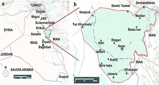
A serosurvey was carried out using cELISA during the period starting from 13.2.2017 to 7.7.2017 to determine the FMD antibodies in apparently healthy small ruminants (sheep and goats) in Garmian region, Kurdistan, Iraq. This region comprises of two districts (Kalar and Kifri) located in north of Iraq which comprises 9.2% of KRG area (Palani, 2018), which consist of 414 villages (Figure 1) and about 585,350 sheep and 87,489 goats are present in the region (Directorate of Veterinay in Garmian, 2017).
Samples analysis
A total of 184 blood samples were collected aseptically from the jugular vein of small ruminant with different ages belong to 18 flocks (flock sizes consist of 250, 195, 200, 150, 150, 180, 130, 350, 185, 100, 200, 660, 120, 430, 400, 225, 80 and 135 heads of sheep and goats) from 14 villages in Garmian region. About 6-8 ml blood was collected by using a vacutainer blood collection tube by jugular vein puncture, then put on ice in a box then let them to coagulate and then transported them to laboratory.
The sera were separated by centrifugation, each serum was put in two Eppendorf tubes which properly labeled then stored at -20°C until examined by ELISA apparatus (BioTeck, USA) for the presence of FMDV antibodies according to Radostits et al. (2007). The kit was provided by ID. vet, Grabels, France, which is a competitive ELISA test for the detection of anti-FMDV non-structural protein (NSP) antibodies in serum and plasma from bovine, ovine, caprine, porcine and all susceptible species.
Statistical analysis
Data were collected and statistically analyzed by using SPSS program (SPSS Inc., Chicago, IL, USA). The chi-square test was employed to calculate the statistical differences between the means.
Result and Discussion
The NSP test is a single test can be used to detect antibodies to any of the seven serotypes of FMD and consider a major advance in the epidemiological tools for FMDV diagnosis (Bronosvoort et al., 2004).
A total of 68 (36.95%) out of the 184 sera samples were seropositive to FMD virus by using NSP-cELISA in Garmian region. According to this study, it is obvious that FMD still endemic and remains a significant disease of sheep and goats. It was predominantly encountered in the cold, dry season (November to March) (Habiela et al., 2010).
The extensive livestock husbandry systems adopted in Garmian region seems to favor conditions for the spread of FMD virus (Habiela et al., 2010). The high rate of the result is due to the fact that sheep and goats in the region are reared under nomadic conditions, and also due to higher livestock mobility in the pastoral system, which facilities high contact and spread of the disease (Mesfine et al., 2019).
According to the results out of 142 sheep sera tested, 56 (39.43%) were positive with no significant differences (p>0.05), compared to 28.57% of goats’ positive samples (Table 1).
Table 1: Infection rate of FMD in sheep and goats according to cELISA results.
| Species | Number of sera tested | Positive result | Prevalence (%) |
| Sheep | 142 | 56 | 39.43 |
| Goat | 42 | 12 | 28.57 |
| Total | 184 | 68 | 36.95 |
P-value=0.135.
The obtained results suggest that certain indigenous animals may have experienced in apparent or subclinical FMD virus infection, and wide prevalence of circulating FMD viruses despite the fact that clinical FMD had not been recorded in villages where the samples were collected as animals repeatedly vaccinated against FMD. The evidence of FMD 3-ABC non-structural protein antibodies in samples from these species may be an indication of natural infection.
Foot-and-mouth disease is spread rapidly within a locality by movement of infected animals to market and by mechanical transmission on items such as clothing, shoes, vehicles, and veterinary instruments (Saleh et al., 2015; Lazarus et al., 2012). The excretion of virus for up to 24 hours prior to the onset of clinical signs means that virus dissemination may have occurred from a flock before any suspicion of disease is raised. This result was agreed with results obtained by Saleh et al. (2015) and Lazarus et al. (2012).
The prevalence of FMD virus according to gender revealed non-significant differences (P˃0.05) in infection rates of female and male animals. Totally among of 155 female animal sera tested, 57 samples (36.77%) were positive, while on contrary, 29 sera samples from male animals tested, 11 (37.93%) were positive (Table 2).
Table 2: Infection rate according to sex based on cELISA results.
| Gender | Number of sera tested | Positive result | Prevalence (%) |
| Female | 155 | 57 | 36.77 |
| Male | 29 | 11 | 37.93 |
| Total | 184 | 68 | 36.95 |
P-value= 0.531
According to age group, the test found that the high rate of positivity (44 %) was at >3 years old with significant differences (P ≤ 0.05) in comparison with low seropositive rate (14.28%) was recorded at <1 year old (Table 3). The age group from 1-3 years exhibited 40% with 26 positive samples out of 65 sera tested.
Seropopositivity was observed high in animals which were sexually mature than immature. The result indicated about one third of sampled sera had FMD seropositive in animals of more than 1 year old. This particular data indicates the extent of recent FMD viral activity in the region’s domestic ruminant population. This might be interpreted as about one-seventh of animals in the region are likely to be affected by an FMD outbreak every year. The result obtained was agreed with previous reports (Mesfine et al., 2019; Hyera et al., 2006).
Table 3: Infection rate according to age groups based on cELISA results.
| Age group (year) | Number of sera tested | Positive result | Prevalence (%) |
| <1 year | 35 | 5 | 14.28 |
| 1-3 years | 65 | 26 | 40 |
| >3 years | 84 | 37 | 44 |
| Total | 184 | 68 | 36.95 |
P-value=0.007
Conclusion and Recommendation
The survey revealed the endemic nature of FMD in sheep and goats in Garmian, as 36.95% of animals were found seropositive for the FMD antibodies. The study results suggested that new future strategy need to be implemented in region to control the increasing incidence of FMD.
Authors Contribution
Authors Hiwa Yassin Essa , Yousef Khalel Bawakhan, Salim Sabah Abdullah, Nisreen Hadi Ali made the serological tests on samples, while Osman M. Jabary Mohammed Tharwat Kareem and Shilan Abubakir Mohammed Amin collected samples and working on publication issues.
Conflict of interest
The authors have declared no conflict of interest.
References


