Journal of Animal Health and Production
Case Report

Parosteal Osteoma in a Green Iguana: A Veterinary Case Report of Medical and Surgical Management
Azlan Che-Amat1*, Asinamai Athliamai Bitrus2, Faez Firdaus Abdullah Jesse1,3, Eric Lim Teik Chung3,4, S. Losheni1, Md Yusoff Sabri5, Muhamad Alif Zakaria6, Nur Asyikin Haron7, Azim Salahuddin Muhamad7, Syahirah Ahmad Affandi7, Yusuf Abba8, Innocent Damudu Peter9, Fitri Wan-Nor1, Idris Umar Hambali10, Bura Thlama Paul1
1Department of Veterinary Clinical Studies, Faculty of Veterinary Medicine, Universiti Putra Malaysia, 43400 UPM, Serdang, Selangor, Malaysia, 2Department of Veterinary Microbiology and Pathology, Faculty of Veterinary Medicine, University of Jos, PMB 2084 Jos, Plateau Nigeria, 3Institute of Tropical Agriculture and Food Security, Universiti Putra Malaysia, 43400 UPM Serdang, Selangor, Malaysia, 4Department of Animal Science, Faculty of Agriculture, Universiti Putra Malaysia, 43400 UPM Serdang, Selangor, Malaysia, 5Department of Veterinary Pathology and Microbiology, Faculty of Veterinary Medicine, Universiti Putra Malaysia, 43400 UPM, Serdang, Selangor, Malaysia, 6Department of Veterinary Laboratory Diagnosis, Faculty of Veterinary Medicine, University Putra Malaysia, 43400 UPM, Serdang, Selangor, Malaysia, 7University Veterinary Hospital, Faculty of Veterinary Medicine, Universiti Putra Malaysia, 43400 UPM Serdang, Selangor, Malaysia, 8Department of Veterinary Pathology, Faculty of Veterinary Medicine, University of Maiduguri, PMB 1069 Maiduguri, Borno Nigeria, 9Department of Theriogenology, Faculty of Veterinary Medicine, University of Maiduguri, PMB 1069 Maiduguri, Borno Nigeria, 10Department of Veterinary Public Health and Preventive Medicine, Faculty of Veterinary Medicine, University of Maiduguri, PMB 1069 Maiduguri, Borno Nigeria.
Abstract | This case report describes the clinical and surgical management of submandibular tumour in a 7 years old female Green Iguana weighing 1.55kg and intensively raised. Physical examination revealed that the iguana had a mass on the ventro-lateral aspect of the head measuring 4 cm x 3 cm in diameter with 7% dehydration. The iguana was placed on lactated ringer solution to correct for dehydration and to stabilize it prior to surgery. Blood samples were collected for complete blood count and serum biochemistry. Impression smear was made from the mass for cytology in addition to radiology diagnosis. Clinical findings based on complete blood count and cytology revealed leucocytosis, lymphocytosis, monocytosis with eosinopenia and presence of clusters of round to spindle shape cells with coarse chromatin embedded in pink cellular matrix. Based on the physical and clinical examination findings, the case was tentatively diagnosed as mesenchymal cell tumour and surgical removal of the tumour was recommended with adequate post-operative care.
Keywords | Iguana, Parosteal Osteoma, Tumour, Medical management, Surgical management.
Received | June 20, 2019; Accepted | September 02, 2019; Published | December 28, 2019
*Correspondence | Azlan Che-Amat, Department of Veterinary Clinical Studies, Faculty of Veterinary Medicine, Universiti Putra Malaysia, 43400 UPM, Serdang, Selangor, Malaysia; Email: c_azlan@upm.edu.my
Citation | Che-Amat A, Bitrus AA, Jesse FFA, Chung ELT, Losheni S, Sabri MY, Zakaria MA, Haron NA, Muhamad AS, Affandi SA, Abba Y, Peter ID, Wan-Nor F, Hambali IU, Paul BT (2019). Parosteal osteoma in a green iguana: a veterinary case report of medical and surgical management. J. Anim. Health Prod. 7(4): 166-170.
DOI | http://dx.doi.org/10.17582/journal.jahp/2019/7.4.166.170
ISSN | 2308-2801
Copyright © 2019 Azlan et al. This is an open access article distributed under the Creative Commons Attribution License, which permits unrestricted use, distribution, and reproduction in any medium, provided the original work is properly cited.
Introduction
The Green iguana (American iguana) scientifically known as Iguana iguana is one of the most common pet reptile. Iguana are arboreal reptiles and their habitat extends from central Brazil to southern Mexico, Bolivia and the Caribbean island (Lazell, 1973; Savage, 2002). Phenotypically, the green iguana possesses a prominent transverse band that spanned from its head to tail and is armed with four powerful limbs. Anatomically, it has a very strong skull compared to the snake but with less mobile jaw. Iguanas can grow big up to 1.5 metre to 1.95-metre-long for an adult and with a life-span of up to 10 years (Simon, 2003; Krysko et al., 2007; O’Malley, 2017). They are ectothermic reptiles where they rely on environment to make themselves warm and have large dewlaps that helps in thermoregulation (Lock, 2006).
Iguanas are large arboreal herbivores reptiles, new-borns and juveniles feed on new leaves, shoots, fruits and blossoms and grasshoppers (Kubiak, 2019a). The adults also feed on vegetation, carrions and birds eggs (Lock, 2006; Krysko et al., 2007). In their natural environment, the adult male Green iguanas have larger territories up to 9,000 m2 than females (Rand et al., 1989). To reach nesting sites, female iguanas travel up to several kilometres, where they nest either communally or alone (Alvarez del Toro, 1960; Rand, 1968; Rand and Dugan, 1983). Nesting sites are usually in sandy open areas, islands, such as riverbanks, or beaches (Burghardt et al., 1977; Campos 2004; Haller and Rodrigues, 2005; Hirth 1963), and females show nesting site fidelity (Bock et al. 1985).
Neoplasm or tumour is an important form of disease in iguanas. In most cases the organs mostly affected are the hematopoietic organs, the liver, and the skin, in addition to tumours or mass of the musculoskeletal system (Hernandez-Diverz, 2003; Lindemann et al. 2017). Baba and Catoi (2007) reported mesenchymal tissue tumours as the most common neoplasm found in reptiles. Mesenchymal tissue tumours are soft tissue or connective tissue tumours that are frequently reported in domestic animals and have a high incidence in some species such as iguanas. This clinical report, describes the clinical, medical and surgical management of a submandibular tumour in a Green iguana.
Clinical Presentation
A 7-year-old female Green iguana weighing 1.55 kg kept as a pet was presented to University Veterinary Teaching Hospital (UVTH) of Universiti Putra Malaysia (UPM) with sub-mandibular mass. Clinical history showed that the iguana was intensively raised, fed with bread and occasionally with vegetables. Four months prior to presentation to UVTH, the Iguana was mixed with other male Iguanas after which she was observed to show aggressive behaviour towards the owner.
Clinical examination revealed that the Iguana was bright, alert and responsive (BAR) but with 7% of dehydration. The moderate dehydration status was evaluated based on the findings of slightly dry and wrinkled skin, sticky mucous membrane and slightly sunken eyes of the Iguana. Further examination revealed a hard mass on the left mandibular region measuring approximately 4 cm x 3 cm in diameter. The pet iguana was immediately placed on intra-coelomic fluid of lactated-ringers’ solution (8 mL) to correct the dehydration status.
Diagnostic Work Plan
Prior to surgical removal of the mass, impression smear of the mass and blood samples were collected for cytology, complete blood count and serum biochemistry. X-ray of the affected area was also taken. Cytology findings revealed a few clusters of round to spindle shape cells with coarse chromatin embedded in pink cellular matrix suggestive of Mesenchymal Cell Tumour (Figure 1). Results from complete blood count revealed the presence leucocytosis due to heterophilia; lymphocytosis and monocytosis with eosinopenia causing leukemoid reaction indicative of systemic infection. Serum biochemistry did not reveal any significant result. Radiograph of the lateral and dorso-ventral aspect of the iguana showed that the lungs and adjoining organs were not affected. From the findings of clinical examination of the iguana the diagnosis made was Mesenchymal cell tumour and surgical removal of the submandibular mass with histopathology analyses was recommended.
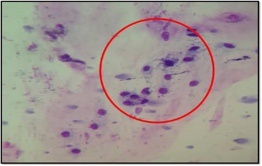
Figure 1: Cytology of the submandibular mass showing a few clusters of round to spindle shape cells with coarse chromatin embedded in pink cellular matrix.
Clinical Management
Anaesthetic protocol and Surgery: Prior to the surgery, the iguana was orally administered Baytril (Enrofloxacin) (5 mg/kg) 1.5 mL x 7/7 and meloxicam 0.2 mL x 3/7. Pre-anaesthetic evaluation of the animal was under the ASA class II (patient with mild disease without substantive functional limitation). Tramadol (5 mg/kg) was administered as an analgesic followed by induction with propofol (3mg/kg). However, because the propofol did not produce the desired effect, the iguana was masked and maintained on 2-4% isoflurane throughout the surgery. Xylocaine and meloxicam 0.2 mg/kg was also topically administered around mandibular surgical site.
The mass site was aseptically prepared with diluted chlorhexidine, iodine and cotton wool. The pet iguana was then placed on dorsal recumbency and the mass located at the left mandible was exposed after draping. Skin incision was made on the lateral edge of the mass using scalpel blade. A blunt dissection was done in between the mass and the muscle layer using a Mayo scissor and necrotic tissues were removed with rat-tooth forceps.
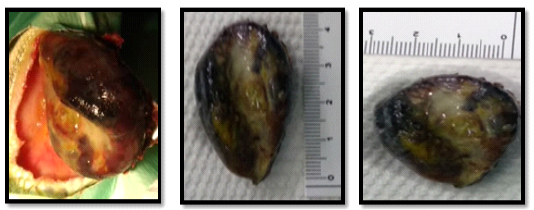
The mass measuring 4 cm x 3 cm was then removed and the necrotic edge of the skin was trimmed (Figure 2). The surgical site was then flushed with normal saline and diluted iodine. The incised skin was sutured using 3-0 Ethilon with horizontal mattress suture pattern and the surgical site was covered with Melolin. Fifteen millilitre (15 mL) of sodium chloride was given intracoelomically to enhance recovery.
Histopathology
Histopathology findings revealed the presence of massive bony mass (osteoid) with minimal narrow space (Figure 3). The bony mass was characterised by the presence of scattered bacteria-like colonies, some with circular pattern and the others found in-between spaces of the bony mass. Additionally, presence of loosely spaced lamellar trabeculae-like structure with outer basophilic and flattened cells were observed (Figure 4). Furthermore, reddish cell (osteoblasts) were also found at the bottom of the bony mass (osteoid). Hence, the condition was tentatively diagnosed as Parosteal Osteoma (Mesenchymal Cell Tumour disease).
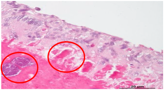
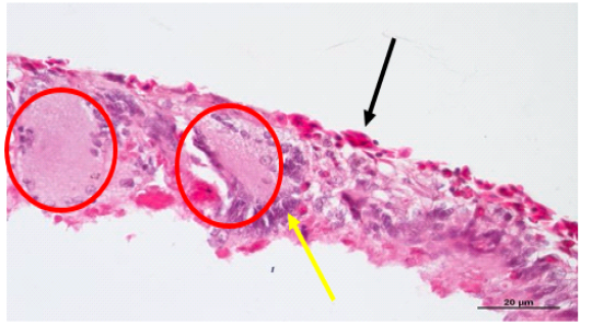
Figure 4: Histopathological findings showing lamellar trabecular-likes structures with outer basophilic and flattened cells.
Post-Operative Therapy and Care
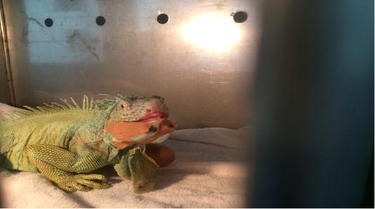
Immediately after surgery (Day 0), the iguana appeared clinically normal (Bright, Alert and Responsive) (Figure 5). The iguana was provided food and a heating lamp to provide thermal support. Additionally, the pet was post-operatively administered with Baytril (Enrofloxacin), 10 mg/kg x 7/7 PO, Tramadol (5mg/Kg) x 7/7 TID, PO and Meloxicam 0.2 mg/kg x 3/7, PO. The suture site was daily flushed with clean sodium chloride solution and monitored three times daily. The iguana was discharged and the client was advised to come for reassessment within 4 to 6 weeks.
Discussion
The iguana has over the years enjoyed tremendous popularity as a zoological exhibit and pet Lizard (Hernandez-Divers, 2003a). Apart from their roles as pets, Iguana helps to improve and maintain forest diversity by eating and dispersing seeds of many plant species. The pet is known to be selective in its diet and consumed seeds have potentially higher germination rates than the uneaten seeds (Krysko et al., 2007). Current improvement in nutrition and captive husbandry has led to several species of these reptiles to survive into adulthood and reaching advanced years (Donoghue, 1996; Hernandez-Divers, 2003b; Kubiak, 2019b).
The goal of the sub-mandibular resection was to alleviate the pain of the Iguana and improvement of it’s welfare. Clinical examination findings and histopathology revealed that the sub-mandibular mass is a mesenchymal cell tumour. Mesenchymal cell tumour is neoplasia of the connective tissue that can either be benign or malignant (Garner et al., 2004). Little is known about the behaviour of this tumour in lizards, however, Hernandez-Divers and Garner 2003b reported that the tumour share similar characteristics as those found in dogs and cats. A few mesenchymal tumours have been reported in reptiles and some are either cartilage or bone tissue tumours that included chondrosarcoma, osteosarcoma and osteochondroma which are predominantly found in Iguana species. Other neoplasm found in reptiles such as iguana, chelonians and snakes include fibro-sarcoma or fibroma and lipoma and also lymphoma or lymphomatoid tumour as the most predominant neoplasm in lizards and snakes (Jacobson et al., 1981; Garner et al., 2004).
It was reported that majority of the tumours commonly encountered in mammals are primarily benign tumours that arise from the mesenchymal elements associated with bone (Hall et al., 2007). According to a survey carried out by Dietz et al. (2015) from 2001 to 2013, thirteen 13 out of 358 clinical cases submitted to veterinary clinics in reptiles were bone tumours. Additionally, the benign bone tumour as well as malignant cartilage and bone tumours reported in the study were mostly found around the head area and limbs of various lizard or iguana species. However, benign neoplasia of the bone are basically rare tumours in reptiles. Hernandez-Diverz (2003) reported several predisposing factors associated with the prevalence of tumours arising in reptiles. The factors include age, immunosuppression, exposure to chronic stressors such as temperature, diet, toxic or radiation, trauma or chronic inflammation. In this clinical case report, the Iguana was 7 years old and has been exposed to other males Iguanas which can lead to aggression and fighting that may cause the trauma.
In tumour therapy, management plays a very crucial role in recovery and healing of the patient. Surgical resection is the recommended and most suitable method for focal masses whose margins are known. Hernandez-Divers and Garner, (2003b) reported that surgical resection was performed on 14/38 (37%) of lizards prior to the diagnosis. The success of the surgical procedure is dependent upon the clinician’s ability to incorporate the standard principles of oncology surgery and reptilian surgical techniques. Alternatively, the use of laser therapy which is less traumatic can be carried out. Radiation therapy and intralesional therapy have been used in snakes with promising results (Done and Mader, 1996; Langan et al., 2001; Hernandez-Diverz, 2003). A medical option available is chemotherapy. However, in contrast with small animal species, chemotherapy is not common in use for treating neoplasia in reptiles except snakes as most of the chemotherapeutic agents must be administered repeatedly through intravenous route and this can be very difficult in iguana species (Straw et al., 1996; Orcutt, 2000; Hernandez-Diverz, 2003). In this clinical case management, diagnosis was achieved based on findings of clinical examination, cytology and histopathology of the excised tumour. In addition, the best treatment option adopted was surgical removal of the superficial mass with adequate post-operative care and this produced a very good result and outcome of the clinical case reported here.
Conclusion
This case report described the medical and surgical management of mesenchymal cell tumour in Green iguana. The case was tentatively diagnosed as Parosteal Osteoma, nonetheless further diagnostic evaluation needs to be carried-out to confirm the type of benign tumour. This type of mesenchymal cell tumour is typically rare in reptiles as malignant tumour is more commonly reported in Iguana than benign mesenchymal tumour.
Acknowledgement
The authors wish to acknowledge the management and staff of the University Veterinary Teaching Hospital, Universiti Putra Malaysia for their technical support during the management of this Veterinary case report.
Conflict of Interest
None to declare.
Authors Contribution
All authors contributed equally and approved the final manuscript.
ReferenceS


