Journal of Animal Health and Production
Case Report

A Protracted Case of Pre-Cervical Uterine Torsion Associated with Fetal Maceration in a Buffalo
Brijesh Kumar1*, Abhishek Kumar1*, R. Sasikala2, Wahengbam Pipelu2, Ishfaq Hajam2, Asif Majid2, Swapna C.R.2, Rohit Kurhe1,Harendra Kumar1
1Division of Animal Reproduction, ICAR-IVRI, Izatnagar, Bareilly-243122, (UP), India; 2Division of Surgery, ICAR-IVRI, Izatnagar, Bareilly-243122, (UP), India.
Abstract | A buffalo in its’ 3rd parity was presented to Referral Veterinary Polyclinic with the history of complete gestation, inappetence for 6-7 days and symptoms of straining and discomfort about 10 days back. The animal was dull and depressed with congested mucous membrane and dry muzzle. Per- vaginal examination revealed only two fingers dilatation of cervix and twisting was felt at the level of 2nd ring, later confirmed by per-rectal examination, which clearly indicated that there was pre-cervical uterine torsion involving cervix. As the anamnesis denoted a delayed case of torsion, uterine adhesions, poor vitality and fragility of uterus was suspected, which could have led to uterine tear if the animal was rolled. Therefore, cesarean section was performed immediately through left lower ventro-lateral oblique incision to relieve the dystocia and a macerated male fetus was delivered. The uterus was cyanotic, fragile and had multiple adhesions with omentum. These adhesions were removed, and the torsion was corrected to certain possible degree. The animal was kept on intensive fluid therapy, antibiotics, anti- inflammatory drugs, oral uterine tonic as well as intrauterine medications for better uterine health and animal recovered uneventfully.
Keywords | Buffalo, Dystocia, Pre-cervical uterine torsion, Uterine adhesions, Fetal maceration
Editor | Asghar Ali Kamboh, Sindh Agriculture University, Tandojam, Pakistan.
Received | January 03, 2019; Accepted | February 03, 2019; Published | February 23, 2019
*Correspondence | Abhishek Kumar, Brijesh Kumar, Division of Animal Reproduction, ICAR-IVRI, Izatnagar, Bareilly-243122, (UP), India; Email: abhiawadhesarita@gmail.com, drbrijeshvet02@gmail.com
Citation | Kumar B, Kumar A, Sasikala R, Pipelu W, Hajam I, Majid A, Swapna CR, Kurhe R, Kumar H (2019). A protracted case of pre-cervical uterine torsion associated with fetal maceration in a buffalo. J. Anim. Health Prod. 7(1): 17-20.
DOI | http://dx.doi.org/10.17582/journal.jahp/2019/7.1.17.20
ISSN | 2308-2801
Copyright © 2019 Kumar et al. This is an open access article distributed under the Creative Commons Attribution License, which permits unrestricted use, distribution, and reproduction in any medium, provided the original work is properly cited.
Introduction
Uterine torsion is a maternal cause of dystocia usually defined as the rotation or spiral twisting of the pregnant uterus on its longitudinal axis (Roberts, 1971). Higher incidence of uterine torsion in buffaloes than cattle is due to anatomical differences like attachment of broad ligaments on the ventro-lateral side of uterus (Noakes et al., 2001), relatively longer and weak musculature of broad ligaments (Ghuman, 2010). The direction of post or pre-cervical torsion can be either clockwise (right) or counter-clockwise (left) (Noakes et al., 2001). Bos indicus cattle is more prone to pre-cervical torsions occurring during the last trimester (Prabhakar et al., 1994; Prasad et al., 2000); while in Bos taurus cattle, cross-bred cattle (56–99%) and buffaloes (87–99%), there is preponderance of post-cervical uterine torsions (Ghuman, 2010; Srinivas et al., 2007).The diagnosis of post cervical uterine torsions by per-vaginal examination and pre-cervical torsions by both per-vaginal and per-rectal examinations becomes essential for making rightful detorsion attempts for correction.
Delayed or long standing cases of torsion (>72 h) is associated with apparent subsiding signs of parturition i.e. shrinkage of udder, loss of teat tumefaction, reabsorption of milk, tightened pelvic ligaments, and reduction in overall perineum relaxation (Srinivas et al., 2007). Limited arterial blood inflow and venous outflow in the twisted uterus leads to ischemia, hypoxia and cell death causing irreversible damage to the endometrium, myometrium and ultimately death of the fetus (Prabhakar et al., 1994). It results in loss of uterine wall elasticity and uterine wall becomes necrosed, fragile and prone to rupture (Noakes et al., 2001). Macroscopically, the color of the uterine wall changes from rose-pink to blue-purple to grey depending upon the degree and duration of torsion (Schönfelder et al., 2007). Bacterial infections and inflammation may spread to dead fetus, fetal fluid, fetal membranes and uterine wall if gained entrance through cervix (Frazer et al., 1996). These inflammatory reactions in uterine wall, tissue anoxia and serosal injury all leads to adhesions of the uterus with the adjoining abdominal viscera or omentum (Ghuman, 2010). Detorsion of the uterus is not possible by rolling the dam after formation of adhesions and laparohysterectomy becomes essential for the correction (Sharma et al.,1995).
History and Clinical Examination
A buffalo in its 3rd parity was presented to the Veterinary Gynecology and Obstetrics section of the Referral Veterinary Polyclinic (Indian Veterinary Research Institute, Izatnagar, Bareilly, U.P) with the history of completed gestation, inappetence for the last 6-7 days, shown signs of straining and discomfort about 10 days back, later diminished. The case was treated locally for a week and there was no significant improvement in the condition of animal. On gross examination, animal was found to be dull and depressed with congested mucous membrane and dry muzzle.
Per vaginal examination revealed only two fingers dilatation of cervix and obstruction or twisting was felt at mid of the cervix,which led to suspicion of the possibility of pre-cervical uterine torsion involving cervix which was later confirmed by per-rectal examination. Torsion was in right side direction as there was tightening of broad ligament of left side and also twisting was well appreciated at the level of cervix near the uterine part. As it was delayed case of torsion, uterine adhesions were suspected, and poor vitality or fragility of uterus could have caused tear of uterus during the process of rolling. Therefore, it was decided to perform cesarean section to relieve the dystocia.
Surgical Correction and Post-Operative Care
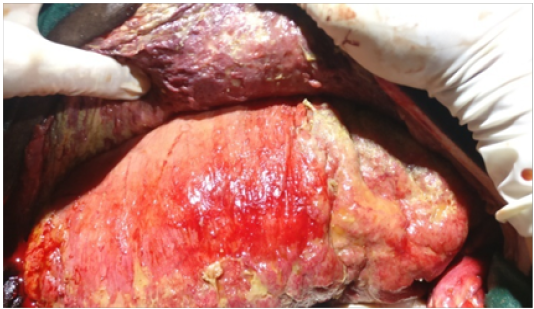
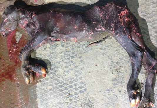
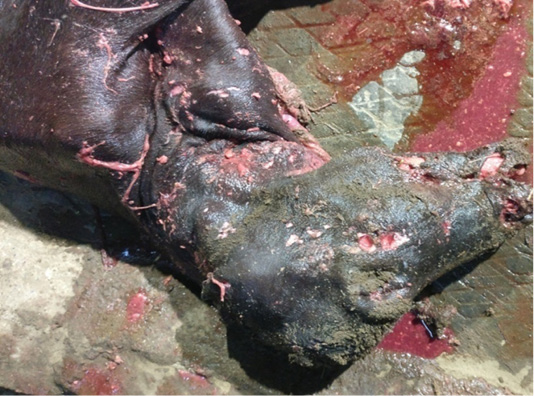
The animal was first stabilized by giving intensive fluid therapy i.e. normal saline (NS) - 5L, dextrose normal saline (DNS)-3L and ringer lactate (RL)-2L over a period of 3 to 5 hours. Antibiotics Enrofloxacin (Quintas®- Intas, India) – 20 mL IM; non-steroidal anti-inflammatory drug Meloxicam (Melonex®- Intas, India) – 10 mL IM, and Pheniramine Maleate (Avil®- Intervet India Pvt Ltd, India) – 10 mL IM were administered along with fluid therapy. Then, the animal was restrained and casted in right lateral recumbency and the incision site (left lower ventro-lateral oblique) was prepared aseptically. Fluid therapy was continued, and incision site was infiltrated by local anaesthetic (2% Lignocaine hydrochloride® inj.) solution. Uterus was necrosed, cyanotic, fragile (Figure 1), and also had multiple adhesions with omentum (Figure 2); as found after skin and muscle incision. Due to this, it was difficult to exteriorize the uterus and incision was made on dorsal curvature of gravid uterus and a macerated male fetus was delivered (Figure 3 and 4). After removing the fetus, easily detachable fetal membranes were removed, and uterine cavity was lavaged with lukewarm normal saline mixed with Povidone iodine®and later Metris® (Metronidazole solution). Uterine incision was closed by absorbable suture material (chromic catgut No. 2 size) using inversion suture pattern (Lembert followed by cushing). Just before the last suture of uterus, Cleanex® bolus (Nitrofurazone I.P 60 mg, Metronidazole I.P 100 mg, Urea I.P 6 gm, Povidone Iodine I.P 60 mg)-4 no., 2 boli in each uterine horn was placed. After suturing of uterus, possible uterine adhesions were removed,and the torsion was corrected to certain possible degree. Abdominal cavity was lavaged with lukewarm normal saline to remove the debris. The peritoneum, muscle layers and subcutaneous tissue were sutured in two layers using vicryl (No. 2 size) in a simple continuous suture pattern. Skin incision was sutured with horizontal mattress suture technique using polyamide (No. 3 size).
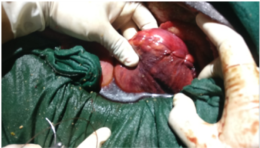
Figure 1: Necrosed and cyanotic uterus
Owner was advised for regular dressing of suture line with povidone iodine® solution and Soframycin ointment. Intensive fluid therapy along with drugs like Quintas®- 20 mL IM sid for 7 days; Melonex®- 10 mL IM sid for 5 days; Avil®– 10 mL IM sid for 5 days; oral herbal uterine cleanser Uterotone® (Cattle remedies- India) - 100 ml P.O. bid for 5 days and Cleanex® (Merial, India) 4 boli intrauterine sid four times on alternate days were prescribed. Animal was discharged with instruction to the owner, for regular dressing of wound and get the stitches removed after 15 days post-operation.
Discussion
Incidence of pre-cervical uterine torsions in buffaloes is low i.e.13-15% of all the cases presented (Ali et al., 2011; Amin et al., 2011). In the present case, this type of torsion was present. As it was delayed case of uterine torsion, rectal examination was necessary to rule out uterine adhesions with the other abdominal structures. Adhesions were present in this case as it was not possible to move the hand on the either side of uterus during per-rectal examination (Noakes et al., 2001). Laparohysterectomy was best and only way of relieving torsion in delayed cases due to uterine adhesions and fragility of uterus to avoid unsuccessful attempts in rolling and chances of uterine tear/rupture after correction (Singh et al., 1978).Therefore, Laparohysterectomy or cesarean section was performed as soon as the case was diagnosed.
Fetal maceration ensues in cervical patency due to ascending infections if fetus is not removed or aborted. Fetal maceration was present in this case as earlier reported in buffaloes (Kochar et al., 1996; Honparkhe et al., 2008; Palanisamy et al., 2018). Although the condition was not dictated as earliest, but therapeutic management and removal of macerated fetus was done as early as possible to avoid life threatening condition in dam due to septicaemia or toxaemia caused by uterine infections.
Acknowledgements
Authors would like to thank Director, Indian Veterinary Research Institute and Incharge, Referral Veterinary Polyclinic for extending the facility for veterinary assistance with the case.
Conflict of Interest
The authors declare no conflict of interest.
Authors contribution
All authors contributed equally during handling of the case. Brijesh and Abhishek contributed to the conception and preparation of the manuscript. Harendra Kumar overall supervised the case and critically guided us during the whole procedure.
References


