Advances in Animal and Veterinary Sciences
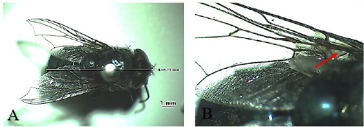
Chrysomya bezziana A. Dorsoventral view; B. Stem vein with bristle (red arrow).
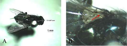
Lucilia sericata; (A) Dorsoventral view; (B) Stem vein without bristle (red arrow).
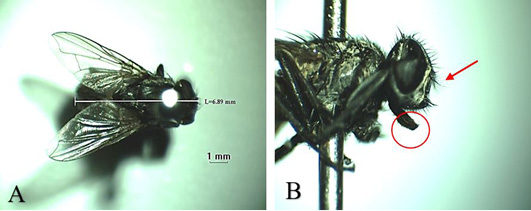
Musca domestica A. Dorsoventral view; B. Arista antennae on dorsal and ventral (red arrows) and licking mouth type (red circle).
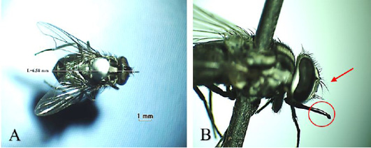
Stomoxys calcitrans (A) Dorsoventral view; (B) Arista antennae only on the dorsal (red arrow) and sucking-piercing mouth type (red circle).
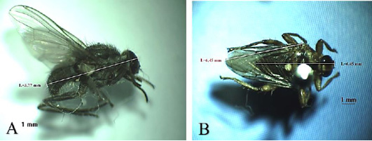
(A) Haematobia exigua lateral view; (B) Hippobosca equina dorsoventral view.
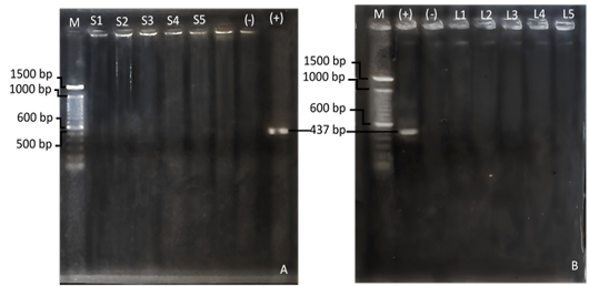
(A) Nested-PCR examination results of the 4th extraction milk sample; (B) nested-PCR examination results of the 4th pooling nuisance fly sample; M (marker); (+) (positive control) C. burnetii strain Nine Mile; (-) (negative control/ aquabidest); S (milk sample); L (fly sample).


