Advances in Animal and Veterinary Sciences
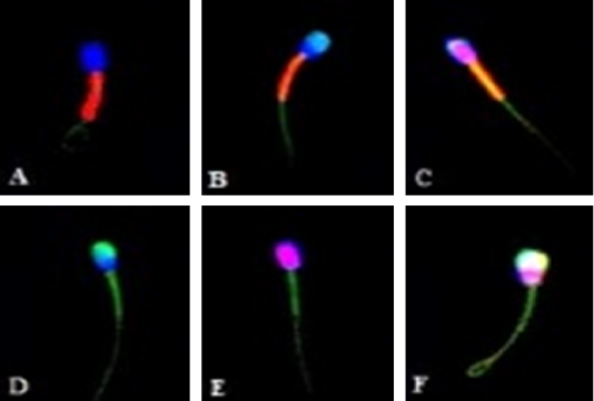
Canine sperm under a confocal laser scanning microscope (600x magnification) after staining with H324, PI, FITC-PSA, and JC-1. (A) Intact plasma and acrosome membrane, and high mitochondrial membrane potential. (B) Intact plasma membrane, damaged acrosome membrane, and high mitochondrial membrane potential. (C) Damaged plasma membrane, intact acrosome membrane, and high mitochondrial membrane potential. (D) Intact plasma membrane, damaged acrosome membrane, and low mitochondrial membrane potential. (E) Damaged plasma membrane, intact acrosome membrane, and low mitochondrial membrane potential. (F) Damaged plasma and acrosome membrane, and low mitochondrial membrane potential.


