Advances in Animal and Veterinary Sciences
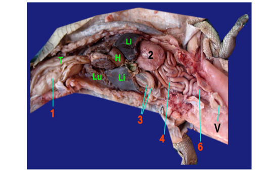
Photograph showing the gastrointestinal tract of crocodile.
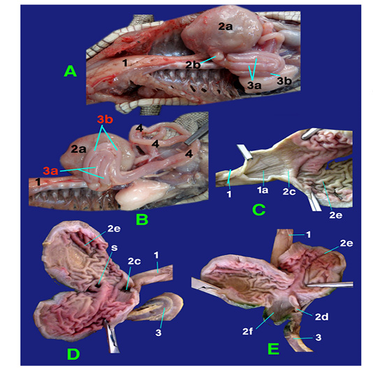
Photograph showing structures of esophagus and stomach.
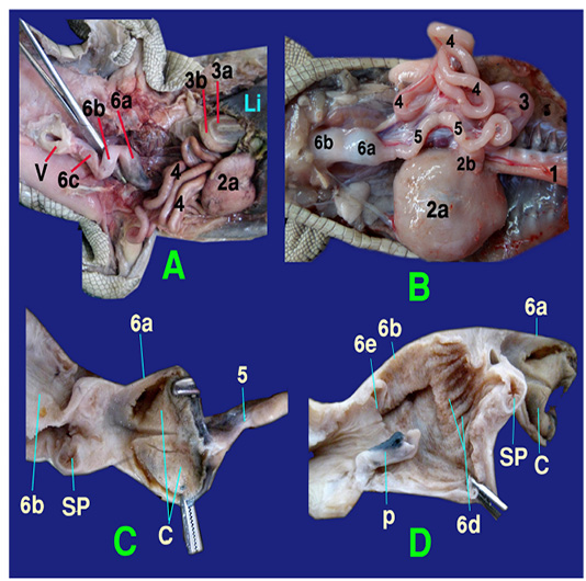
Photograph showing the structures of colorectum
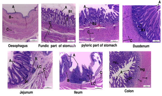
Photograph showing the histology of gastro-interstinal tract.
A, tunica mucosa ; B, tunica submucosa; B1, fundic gland; B2, pyloric gland; B3, duodenal gland; gc, goblet cell ; LC, lymphocyte; mg, mucous gland.
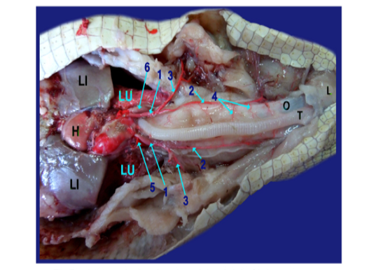
Photograph showing the origin of the right and left aorta.
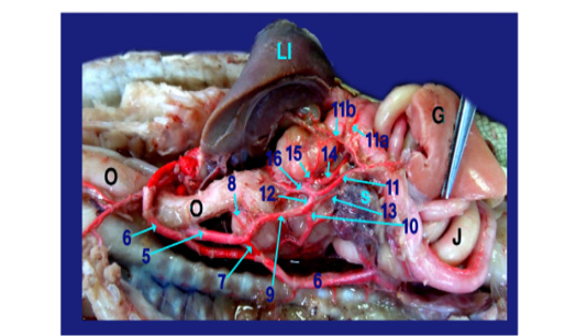
Photograph showing the branches of celiacomesentric artery.
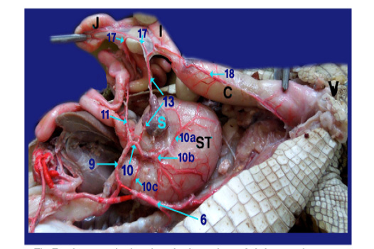
Photograph showing the branches of right gastric artery.
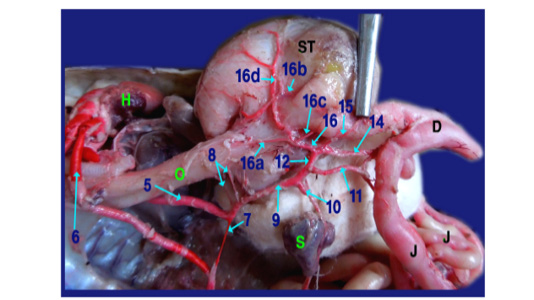
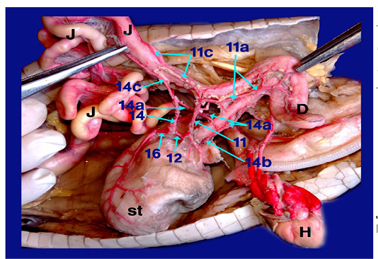
Photograph showing the branches of gastroduodenal artery.
Photograph showing the arterial supply of duodenum and jejunum.


