Advances in Animal and Veterinary Sciences
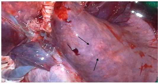
Congested and consolidated pneumonic lung with dark red discoloration (arrow)
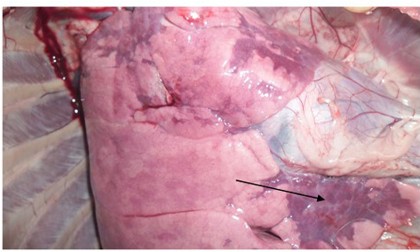
Congestion and consolidation of cardiac lobe of lung (Arrow)
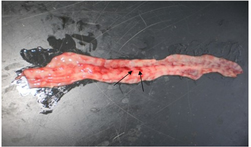
Zebra striping in colon
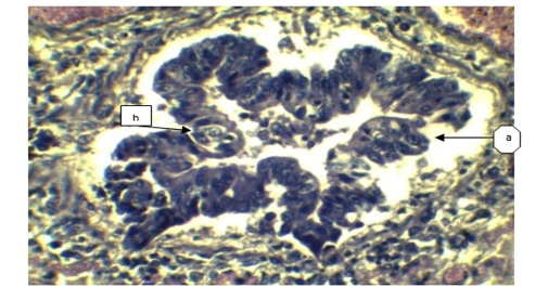
a) Sloughing off surface epithelium of bronchi; b) Necrosis of bronchiolar epithelium. (H&E x 40).
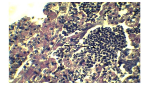
Infiltration of inflammatory cell in Lung parenchyma (Arrow), feature of interstitial pneumonia. (H&E x 40)
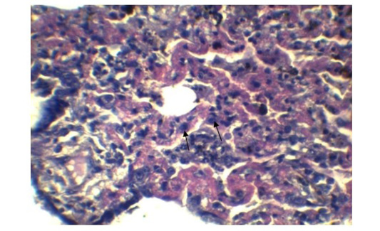
Expanded alveolar wall due to prominent type-II pneumocytes. (H&E x 40).
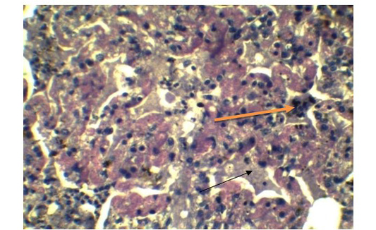
Presence of syncytial cell in the alveoli (Yellow arrow). Presence of fibrin exudates in parenchyma (Black arrow) (H&E x 40).
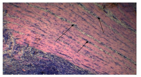
Inflammatory cells in the submucosa (Arrows). (H&E x 10).
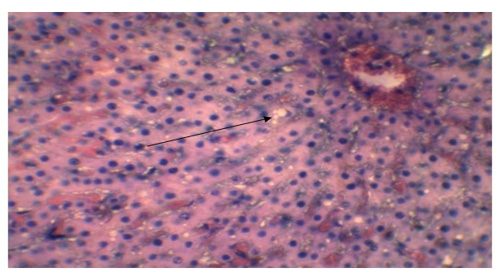
Vacuolar or fatty degeneration in the cytoplasm of hepatocyte (Arrow). (H&E x 40).


