South Asian Journal of Life Sciences
Dark black necrotic with severe myositis and hemorrhages in thigh muscles of Swiss albino mice infected with Clostridium chauvoei.
Inflammation, dark black of heart muscles infected with Clostridium Chauvoei in Swiss albino mice.
Dark black and greenish color due to necrosis and hepatomegaly due to infection of Clostridium Chauvoei in liver of Swiss albino mice.
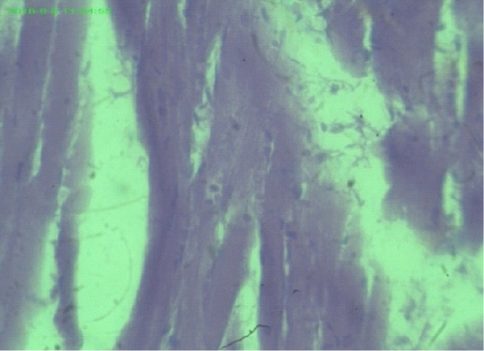
Uniform and dense striated muscles of thigh and intramuscular gases accumulate (arrows), (H&E staining at 40x magnifications).
Myocardial fiber attenuation and separation, disarray of myofibrils (arrows), (H&E staining at 40x magnifications).
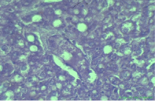
Edematous, swollen hepatocytes and multifocal hepatic necrosis lesions (arrows), (H&E staining at 40x magnifications).
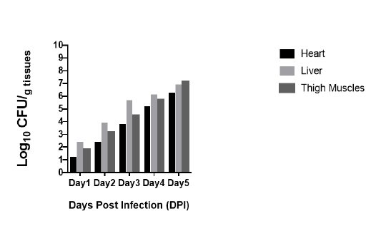
Bacterial load in heart, liver and thigh muscles after inoculation of Clostridium Chauvoei in mice 0.1ml (1x10⁻⁵) CFU/ml. Bacteria are expressed as log10 number of CFU/gram of heart, liver and thigh muscle.


