South Asian Journal of Life Sciences
Case Report
South Asian Journal of Life Sciences. 1 (1): 19 – 20Caseous Lymphadenitis in a Goat
Faez Firdaus Jesse Abdullah1,3*, Abdinasir Yusuf Osman1,2, Lawan Adamu1,5, Nurul Alia Azri1, Abdul Wahid Haron1,3, Mohd Zamri Saad2, Abdul Rahman Omar2,4 , Abdul Aziz Sharee1
- Department of Veterinary Clinical Studies
- Department of Veterinary Pathology and Microbiology
- Research Centre for Ruminant Disease, Faculty of Veterinary Medicine, Universiti Putra Malaysia, 43400 UPM Serdang, Selangor, Malaysia
- Institute of Bioscience, Universiti Putra Malaysia, Malaysia
- Faculty of Veterinary Medicine, University of Maiduguri, PMB1069, Borno State, Nigeria
*Corresponding author:jesse@putra.upm.edu.my
ARTICLE CITATION:
Abdullah FFJ, Osman AY, Adamu L, Azri NA, Haron AW, Saad MZ, Omar AR and Sharee AZ (2013). Caseous Lymphadenitis in a Goat. South Asian J. Life Sci. 1 (1): 19 – 20.
Received: 2013–07–10, Revised: 2013–11–30, Accepted: 2013–12–04
The electronic version of this article is the complete one and can be found online at
(
http://nexusacademicpublishers.com/table_contents_detail/25/143/html
)
which permits unrestricted use, distribution, and reproduction in any medium, provided the original work is properly cited
ABSTRACT
Despite the sero–positivity of caseous lymphadenitis (CLA) in Malaysian farms, cases with clinical signs of CLA are rarely encountered. An 8–month–old Saanen breed of goat was reported with a history of a subcutaneous mass beneath the jaw, on the right side. The consistency of the mass was non–movable, solid and hard on deep palpation. A tentative diagnosis of caseous lymphadenitis (CLA) was made based on the characteristic of the lesions presented and its contagious nature. The mass was incised and the purulent aspirate showed growth of Corynebacterium pseudotuberculosis on culture. Agar gel precipitation test (AGPT) was positive for Corynebacterium pseudotuberculosis. The mass was lanced and routine wound flushing resulted in recovery.
Caseous lymphadenitis (CLA); a chronic contagious disease of sheep–goat populations worldwide has considerable economic consequences. The direct effects of CLA include loss of fertility, loss of draught power, retardation of growth, gradual emaciation and condemnation of carcasses, while indirect losses can be attributed to the disruption in trade of animals and derivative products (Williamson, 2001).
CLA is caused by a bacterium known as Corynebacterium pseudotuberculosis. The entry to the host is mainly through non–intact skin such as open wounds and abrasions (Fontaine and Baird, 2008) and also mucous membrane (Windsor, 2011). However, other routs have been described in detail by Jesse et al. (2013) and Adza Rina et al. (2013).
Transmission occurs from environmental contamination. Subsequently, the organism migrates within the phagocytes to the local lymph nodes and internal organs, causing inflammatory reactions. Repeated cycles of bacterial replication, followed by necrosis, subsequent death of inflammatory cells forms the characteristic of abscess of superficial lymph nodes to which the disease is clinically characterized (Windsor, 2011).
An eight month old female Saanen breed goat was presented for evaluation of large subcutaneous mass at the base beneath the jaw, on the right side. Vaccination and deworming status was unknown. Two weeks prior to the visit, the owner noticed 4 out of 60 goats in the herd had mass growing around the submandibular and pre–scapular region. There was no history of trauma, injection or other potential inciting cause for development of the mass. However, the total herd (n=60) were imported from Australia approximately three weeks prior our visit to the farm. The affected ones were isolated from the rest of the herd. The farm was designed as a dairy goat farm producing milk commercially for only local consumption.
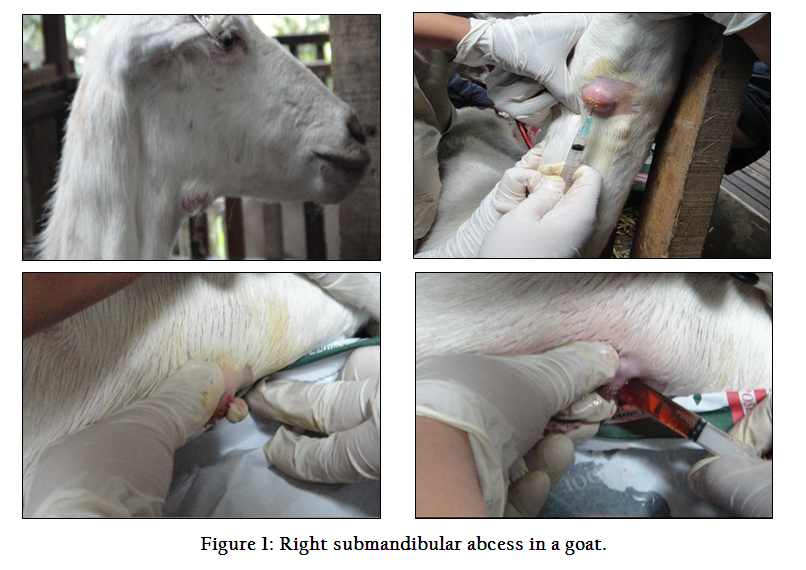
Physical examination reveals vital parameters were within normal range. Body condition score was 2.5/5. There was enlargement of the right submandibular lymph node. The site of enlargement was alopecic; the consistency was non–movable, solid and hard. Despite the clinical lesions, the goat’s appetite, bowel and urination were normal. The same observation was recorded in the other three goats with which similarity, in terms of clinical lesions, exists.
A tentative diagnosis of caseous lymphadenitis was made based on the history and characteristic of the lesions. The area was aseptically prepared and a 21–gauge, 3.0–cm over–the needle catheter inserted into the mass; 2 mL of purulent exudate was aspirated and submitted bacteria cultures. Microscopic examination of the Gram–stained smear revealed the presence of Gram–positive pleomorphic rods. Bacterial culture results revealed growth of C. pseudotuberculosis, which confirmed the diagnosis.
Management of the case involved establishing drainage and instituting daily lavage of the abscess with diluted hibiscrub (chlorhexidine) and iodine solution. Orospray (sulphanilamide, chlortetracycline) was applied on the lesion as a topical antibiotic. Wound sarex was finally applied around the lesion for the purpose of avoiding environmental contamination and insect agitation.
In many cases, abscessation caused by C. pseudotuberculosis in small ruminant flocks (sheep and goats) present at the point of entry into the skin or in a nearby lymph node. Abscessation involving other soft tissues has also been reported. In internal form of CLA infection, vital organs are more likely to develop abscessation than any other organs (Abdinasir et al., 2012).
Treatment of abscesses caused by C. pseudotuberculosis involved drainage and lavage of the abscess in combination with antimicrobial therapy including penicillin, doxycycline, trimethoprim–sulfonamide or a combination of these (Beck et al., 2011). Recurrence of the disease even after removal of abscess is more likely to occur. In addition, presences of internal abscesses are also a major constraint for effective therapy (Dorella et al., 2006). Control and prevention is the key to prevent spread of the disease. In conclusion, this case was diagnosed and treated employing routine methods.
ACKNOWLEDGMENT
The authors would like to thank Mohd Jefri Norsidin for referral and ongoing management of the case, and the staff of the Department of Pathology and Microbiology, Fcaulty of Veterinary Medicine, Universiti Putra Malaysia (UPM), in particular, Mohd Azri Roslan for processing the samples.
CONFLICT OF INTEREST
The authors declare that they have no conflict of interest with the contents of this paper in any respect.
REFERENCES
Abdinasir YO, Jesse FFA, Saharee AA, Jasni S, Khairani–Bejo S, and Haron AW (2012). Clinico–Pathological Changes in Mice Following Experimental Infection with Whole Cell and Exotoxin (PLD) Extracted from C. pseudotuberculosis. Journal of Animal and Veterinary Advances, (11): 4064–4072.
Adza–Rina MN, Zamri–Saad M, Jesse FFA, Saharee AA, Haron AW and Shahirudin S (2013). Clinical and pathological changes in goats inoculated with Corynebacterium pseudotuberculosis by intradermal, intranasal and oral routes. Online Journal of Veterinary Research, (17): 73–81.
Beck A, Baird JD, and Slavic D (2011). Submandibular lymph node abscess caused by Actinomyces denticolensin a horse in Ontario. Canadian Veterinary Journal (52): 513–514. PMid:22043071 PMCid:PMC3078004
Dorella FA, Pacheco L G C, Oliveira SC, Miyoshi A, and Azevedo V (2006). Corynebacterium pseudotuberculosis: microbiology, biochemical properties, pathogenesis and molecular studies of virulence. Veterinary Research. (37): 201–218.
http://dx.doi.org/10.1051/vetres:2005056
PMid:16472520
Fontaine MC and Baird GJ (2008).Caseous lymphadenitis. Small Ruminant Research, (76): 42–8.
http://dx.doi.org/10.1016/j.smallrumres.2007.12.025
Jesse FFA, Adamu L, Abdinasir YO, Ahmad Fauzan M, Abd Wahid H, Abdul Aziz S. et al. (2013). Polymerase Chain Reaction Detection of C. pseudotuberculosis in the Brain of Mice Following Oral Inoculation. International Journal of Animal and Veterinary Advance (5): 29–33.
Williamson LH, (2001). Caseous lymphadenitis in small ruminants. The Veterinary Clinics of North America. Food Animal Practice, (17): 359–71.
PMid:11515406
Windsor P A (2011). Control of caseous lymphadenitis. The Veterinary Clinics of North America. Food Animal Practice, (27):193–202.
http://dx.doi.org/10.1016/j.cvfa.2010.10.019
PMid:21215903