Journal of Infection and Molecular Biology
Research Article
Journal of Infection and Molecular Biology 2(1): 1 – 5Protein Profile of Normal and Hydropericardium Infected Poultry Liver Using Sodium Dodecyl Sulphate–Polyacrylamide Gel Electrophoresis and Western Blotting
Shahid Khan1*, Shamsher Ali Khan1, Sayyed Rahimullah Shah1, Faizul Hassan1, Kaleem Allah1, Khasro Kaleem2
- Microbiology Section, Veterinary Research and Disease Investigation Centre, Swat, KPK province, Pakistan
- Livestock Research and Development Station Dir (Lower), KPK province, Pakistan
*Corresponding author: wazirkhattak@yahoo.com
ARTICLE CITATION:
Khan S, Khan SA, Shah SR, Hassan F. Allah K, Kaleem K (2014). Protein profile of normal and Hydropericardium infected poultry liver using sodium dodecyl sulphate–polyacrylamide gel electrophoresis and western blotting. J. Inf. Mol. Biol. 2 (1): 1 – 5.
Received: 2013–04–26, Revised: 2013–09–27, Accepted: 2013–10–01
The electronic version of this article is the complete one and can be found online at
(
http://dx.doi.org/10.14737/jimb.2307-5465/2.1.1.5
)
which permits unrestricted use, distribution, and reproduction in any medium, provided the original work is properly cited
ABSTRACT
Preparation of autogenous vaccine against Hydropericardium syndrome (HPS) and subsequent administration to chicks is a common practice in Pakistan. However, due to lack of quality control testing and evaluation procedures, the accurate quantity of virus from a given clinical sample of liver always remains questionable. Therefore, the present study has been conducted to differentiate the protein profile of normal and HPS infected liver lysate with respect to the availability of immunogenic proteins using sodium dodecyl sulphate–polyacrylamide gel electrophoresis (SDS–PAGE) and western blotting techniques. Using commercially available vaccine against HPS (Ottomans Parma), hyperimmune serum was raised in immunologically naïve broiler birds (3–weeks old, n = 60) and subjected to agar gel precipitation test (AGPT) for screening HPS positive serum samples. Of the total examined birds, only five birds were found positive. The commercially available HPS autogenous vaccine, liver from HPS positive bird and normal liver samples were subjected to protein extraction and measurement. SDS–PAGE analysis revealed 15–structural polypeptide bands in both HPS infected poultry liver and autogenous vaccine. Contrary to this, 10–structural polypeptide bands were observed in normal liver. The polypeptides of HPS autogenous vaccine and both normal and HPS infected livers, separated on 9% gel were transferred onto nitrocellulose membrane. The western blot analysis of proteins from Hydropericardium virus infected liver and HPS autogenous vaccine showed one immunogenic protein of molecular weight 15-20 kDa These differences could be used for identification and differentiation between clinically diseased and healthy birds as well as vaccinated by autogenous vaccine.
INTRODUCTION
Hydropericardium syndrome (HPS) was first recognized in broiler–rearing area of Angara Goth, Karachi in 1987 (Muneer et al., 1989). The disease was thought to be a multifactorial syndrome initially, which might have occurred either due to bacterial infection, poisoning with disinfectant, insecticides (McCune et al., 1962) or toxic fat (Sanger et al., 1958). Nevertheless, the Koch’s postulates, from liver homogenate of the infected birds, have been proved by a number of researchers (Cheema et al., 1989; Muneer et al., 1989). Now, Fowl adenovirus serotype–4 (FAdV–4) has been considered the cause of HPS in birds (Afzal and Ahmed 1990).
Since 1989 autogenous vaccine was prepared from infected liver suspension to protect the broiler flock from HPS. However, the protection provided by this vaccine is not long-lasting and disease has been reported even in vaccinated birds (Afzal and Ahmad., 1990). Though the disease can be diagnosed from gross lesions and histopathological changes in the liver, various serological tests such as agar gel immunodiffusion (AGID), counter immuno–electrophoresis (CIE) indirect hemagglutination and ELISA (Chandra et al., 2000) has been found useful.
Without known quality control procedures, many commercial labs in Pakistan are manufacturing autogenous liver homogenate vaccine. As the quantity of the virus is not measured, some of the livers used in making the vaccine may not either have sufficient concentration of virus or no virus at all. The present study, therefore, has been conducted to differentiate the protein profile of normal and HPS infected liver lysate with respect to the availability of immunogenic proteins using SDS–PAGE and western blotting technique. The method applied could be used for identification and differentiation between clinically diseased and healthy birds as well as to prepare autogenous vaccine.
MATERIALS AND METHODS
Based upon clinical history and necropsy lesions suspected of HPS, infected liver samples (n = 30) were collected aseptically from poultry farms in and around district Lahore. The normal liver samples were also collected from clinically healthy birds at UVAS Lahore as a negative control. The samples were transported at 40C and stored at –200C for further use. Three types of autogenous vaccines and one cell cultured vaccine against HPS virus used in the analysis were purchased from local market at Lahore, Pakistan.
Preparation and evaluation of antibodies against HPS virus
Sixty chicks (3 week old) were used to raise antibodies against HPS virus. Based upon four type of commercially available HPS vaccine, the birds were divided into four groups and given each type of vaccine. The birds were bled on 24th day; serum was collected and stored at –20 °C until used. The oil based cell culture HPS vaccine (Rhone Merieux, (USA)) was used as an antigen in agar gel precipitation test (AGPT) (Kumar et al., 1997).
Sodium dodecyl polyacrylamide gel electrophoresis (SDS–PAGE)
Sodium dodecyl polyacrylamide gel electrophoresis (SDS–PAGE)
To make component peptides for use in SDS–PAGE, the positive liver samples (tested by AGPT) were homogenized and subjected to sonication by giving 10 shots at the interval of five minutes each using sonicator (EILA, Japan). Liver samples were processed by chloroform treatment method. Chloroform was added to the supernatant in 2:1 ratio, shaken slowly for 15 minutes manually and then subjected to sonication again. The sonicated samples were placed in centrifuge machine (Merck, Japan) for 15 minutes at the speed of 4,000 rpm. After centrifugation, the supernatant was taken in a sterile microfuge tube and subjected to ammonium sulphate precipitation (Pal et al., 2004). Similarly, the normal liver and HPS autogenous vaccines were processed. A stock solution containing bovine serum albumin (100mg/mL) was prepared in normal saline which was further diluted to prepare concentrations of 1, 20, 40, 60, and 80 88 mg/mL.
Using bovine serum albumin as standard, the UV spectrophotometer (at 280 nm wavelength) was used to calculate protein concentrations in diseased, healthy and vaccine samples (Kumar and Chandra, 2004). Working solution for loading gel was prepared as described previously (Maiti and Sarkar, 1997). Unstained protein markers (Fermentas, USA) were used. Samples polypeptides were separated using a distinctive gel of 10% acrylamide composition to analyze the entire profile of supernatants. A 30 mL of gel–mixture was prepared by mixing already prepared monomer stock (30%) (9 mL), resolving gel buffer (31.6%) (7.5 mL), 10% SDS (300 µL), ammonium persulphate 10% (150 µL) and TEMED (12 µL) and water (12 mL). While the stacking gel (6%) was prepared to a total quantity of 10 mL. The electrophoresis tank was filled with 6L of tank buffer (Electrophoresis buffer 10%) and the casting stand (containing gel) was placed vertically in electrophoresis tank. Finally, 30 µL of denatured protein samples were loaded in each well and the gel was run at voltage 4.54V/cm. The power was turned off when dye border reached near the gel bottom. Gel was removed from the cassette and placed in a staining dish containing deionized water. After a quick rinse, the water was poured off and the stain was added. Coomassive brilliant blue dye (0.1%) in 50% methanol was used and the protein bands in polyacrylamide gel were detected in 10% glacial acetic acid. Gel was agitated for 3–4 hours, stained in acetic acid and methanol to remove the extra dye.
Identification of immunogenic proteins using Western Blotting technique
The Bio–Ice cooling unit (BIO–USA) was filled with water and stored in freezer at –85 °C until used. The transfer buffer (5%) was chilled to 4 °C The nitrocellulose membrane (0.22 μm pore size) and the filter paper were cut to the dimensions of the gel (containing the fractionated protein).The gel sandwich was prepared as described by Kumar and Chandra (2004). The cassette containing sandwich was placed in module (Bio–Red, USA) in such a way that the gel was at cathode side and nitrocellulose membrane on anode side. The frozen Bio–Ice cooling unit was added, placed in tank (Bio–Red, USA) and the tank was completely filled with transfer buffer (5%). The blot was run at 100 volts for one hour. Upon completion of the run, the blotting sandwich was disassembled and the membrane was removed.
A duplicate of western blot was stained for visualization of the bands. The blot was stained using working stain (2%) to visualize protein bands on nitrocellulose. The blots were differentiated in running tap water. Later the stain was completely removed by washing under tap water. The unstained western blot was incubated overnight in blocking buffer (3%) at room temperature to block non-specific sites on membrane. The blotting paper was washed 3 times in washing buffer (0.5%) for 10 minutes each to remove unbound blocking agent. Filter paper (Whatman, 3mm) and blotting paper, both were cut into equal size using a clean blade. The filter paper was saturated with serum diluted in blocking buffer (3%) (1:4) assembled with blotting membrane having protein bands and were sealed by clumping between two glass plates .The sandwich was incubated at room temperature for 45 minutes. The filter paper was removed and blotting membrane was washed three times by shaking in 100 mL/wash, in PBS–Tween 20 solution (washing buffer) for 5 minutes each at room temperature. The horseradish peroxidase conjugated anti–chicken IgG was diluted in 10 mL blocking solution (3%) (1:50). The filter paper was saturated with diluted horseradish peroxidase conjugated anti–chicken IgG and were sealed by clumping between two glass plates. The sandwich was incubated at room temperature for 45 minutes. The filter paper was removed and blotting membrane was washed three times by shaking in 100 mL/wash, in washing buffer for 5 minutes each, at room temperature.
Noble agar (0.05 gm) was dissolved in 50 mL deionized water by gentle heating. Four mL of substrate was mixed with four mL of melted agar and poured slowly on blotting membrane to prevent bubble formation. Also the filter paper was fully saturated in 5 mL substrate solution and placed on blotting membrane. Results were recorded within 15 minutes.
RRESULT
Sixty broilers were divided into four groups viz A, B, C and D (n = 15 each). Three different liver homogenate vaccines (A, Band C) and one oil based cell culture vaccine (D) (Rhone Merieux, Georgia) was used for raising hyper immune serum against HPS virus. The birds were kept for screening of antibodies against HPS virus before vaccination. The birds were immunized with vaccinal protein concentration (30mg/mL) at the dose rate of 0.5 mL/bird at 1st day of vaccination 0.75 mL/bird on 7th day and 1 mL/bird on 14th day. Serum was collected at 9th day after 2nd boosting. All serum samples were then subjected to Agar gel precipitation test (AGPT). Serum raised in response to oil based cell culture HPS vaccine produced precipitation line.
The homogenates of HPS infected livers (20%) collected from different poultry farms were screened by hyperimmune serum raised against oil based cell culture HPS vaccine using AGPT. Precipitation lines were observed in three liver samples. The positive samples were stored at –4°C till further used. Absorbance of the sample by spectrophotometer was used to determine the concentration of protein samples from the calibration curve (Table 1). A standard curve was created by plotting concentration of the standard versus regression analysis of A280, as shown in the Figure 1. The polypeptides of normal and HPS infected livers were resolved by SDS–PAGE using discontinuous buffer system. In 9% acrylamide gel, normal liver and HPS autogenous vaccine–1 yielded 10 polypeptides while HPS infected liver and HPS autogenous vaccine–2, yielded 15 polypeptide bands which were visualized on Coomassie brilliant blue R–250 staining (Figure 2).
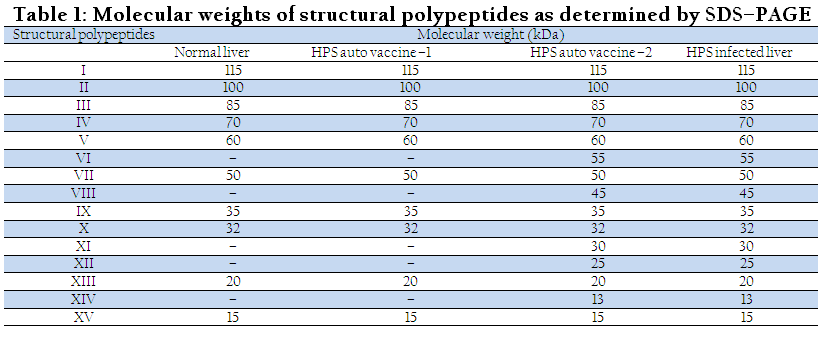
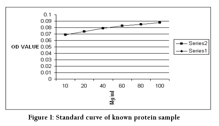
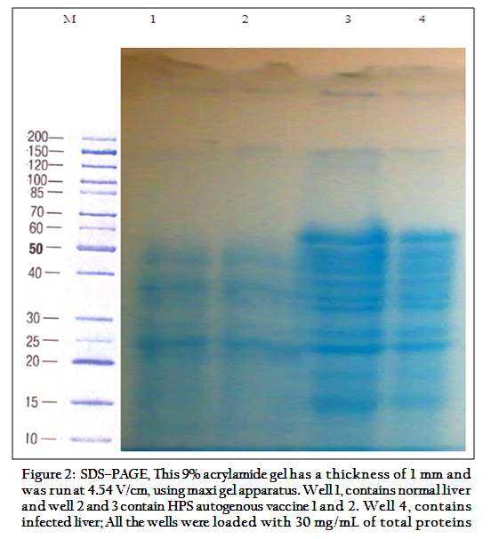
Figure 2: SDS–PAGE, This 9% acrylamide gel has a thickness of 1 mm and was run at 4.54 V/cm, using maxi gel apparatus. Well 1, contains normal liver and well 2 and 3 contain HPS autogenous vaccine 1 and 2. Well 4, contains infected liver; All the wells were loaded with 30 mg/mL of total proteins
The polypeptides of HPS autogenous vaccine, normal and HPS infected livers, separated on 9% gel were transferred onto nitrocellulose membrane (0.22 μm) by Western blotting technique (Kumar and Chandra, 2004). An immunogenic band was detected in well containing HPS virus infected liver homogenate and HPS autogenous vaccine (Ottoman Pharma, Lahore) in the form of color spot. The position of spot was compared with stained SDS–PAGE gel. It was found to be between 15–20 kDa both in HPS infected and HPS autogenous vaccines (Figure 3)
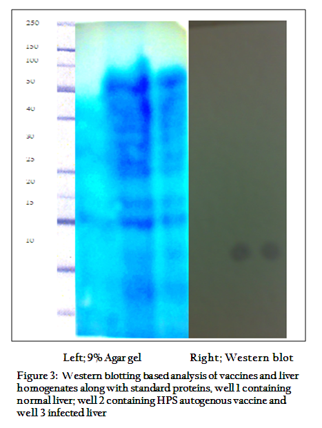
Figure 3: Western blotting based analysis of vaccines and liver homogenates along with standard proteins, well 1 containing normal liver; well 2 containing HPS autogenous vaccine and well 3 infected liver
DISCUSSION
In the present study, the infected liver and HPS autogenous vaccine revealed 15–polypeptides and one immunogenic band which is different from previous observations made by number of researchers. For example, Maiti and Sarkar (1997) have verified 11 polypeptides, ranging in molecular weights from 10 to 100 kDa in FAV serotype 1. Nazerian et al. (1991) reported 11 structural polypeptides in FAV serotype 2 that ranged in molecular weight from 97 to 14 kDa. Li et al. (1999) reported at least 14 polypeptides in chicken embryo lethal orphan (CELO) virus (fowl adenovirus type 1). Kumar and Chandra (2004) observed a total of 12 polypeptides ranging in molecular weight between 13.8KDa and 110.0 kDa and also seven immunogenic polypeptides ranging in molecular weight between 15.8 and 110.0 kDa.
Contrary to this, normal liver revealed 10–polypeptides with a difference of 5–polypeptide and one immunogenic band from diseased or infected liver. The present study revealed that some HPS autogenous vaccine may not contain even single immunogenic proteins and thus, may not be able to provoke an effective immune response. The local manufacturer should take into consideration the fact and appropriate procedures should be adopted such as described here; evaluation of vaccine using SDS PAGE and western blotting along with normal and diseased clinical samples.
REFERENCES
Afzal M, Ahmad I (1990). Efficacy of an inactivated vaccine against HPS in broilers. Vet. Rec. 126(3): 59 – 60.
PMid:2301129
Chandra R, Shukla SK, Kumar M (2000). The Hydropericardium syndrome and Inclusion Body Hepatitis in domestic fowl. 321(2): 99 – 111.
Cheema AH, Afzal M, Ahmad J (1989). An adenovirus infection of poultry in Pakistan. Rev. Sci. Tech. Off. Int. Epiz. 8(3): 789 – 795.
Kumar R, Chandra R (2004). Studies on structural and immunogenic polypeptides of hydropericardium syndrome virus by SDS–PAGE and western blotting. Comp. Immunol. Microbiol. Infect. Dis. 27(3): 155 – 161.
http://dx.doi.org/10.1016/j.cimid.2003.08.003
PMid:15001310
Kumar R, Chandra R, Shukla SK, Agrawal DK, Kumar M (1997). A preliminary study on the causative agent and control of the disease by inactivated autogenous vaccine. Comp. Immunol. Microbiol. Infect. Dis. 29(3): 55 – 60.
Li P, Bellett AJ, Parish CR (1983). A comparison of the terminal protein and hexon polypeptides of avian and human adenoviruses. J. Gen. Virol. 64: 1375 – 1379.
http://dx.doi.org/10.1099/0022-1317-64-6-1375
PMid:6304239
Maiti NK, Sarkar P (1997). Structural polypeptides of different clinical strains of avian adenovirus type–1. Comp. Immunol. Microbiol. Infect. Dis. 20(1): 53 – 58.
http://dx.doi.org/10.1016/S0147-9571(96)00027-6
http://dx.doi.org/10.1016/S0147-9571(96)00028-8
Mc-Cune EL, Sevage JE, Dell BLO (1962). Hydropericardium and ascites in chicks fed a chlorinated biphexyl product. Poult. Sci. 41: 259 – 299.
Muneer MA, Ajmal M, Arshad M, Ahmad MD, Chaudry ZI, Khan TM (1989). Preliminary studies on HPS in broilers in Pakistan. Zootech. Intl. 5: 46 – 48.
Nazerian K, Lee LF, Payne WS (1991). Characterization of Serotype-11 structural polypeptides in FAV serotype 2. Comp. Immunol. Microbiol. Infect. Dis. 35: 572 – 578.
Pal JK, Godbole D, Sharma K (2004). Staining of proteins on SDS polyacrylamide gels and on nitrocellulose membranes by Alta, a colour used as a cosmetic. Biochem. Biophy. Methods. 61(3): 339 – 347.
http://dx.doi.org/10.1016/j.jbbm.2004.06.008
PMid:15571781


