Research Journal for Veterinary Practitioners
Case Report
Research Journal for Veterinary Practitioners. 1 (2): 20 – 22Clinical Aspects of Canine Distemper in 1.5 Year Old Labrador retriever bitch bit Bitch
Arslan Tariq*, Asim Shahzad, Sarwat Tahira
-
Faculty of Veterinary Sciences, University of Agriculture Faisalabad, Pakistan. Postal code: 38000
*Corresponding author:dr.arslantariq3418@live.com
ARTICLE CITATION:
Tariq A, Shahzad A, Tahira S(2013). Clinical aspects of canine distemper in 1.5 year old Labrador retriever bitch. Res. J. Vet. Pract. 1 (2): 20 – 22.
Received: 2013-07-30, Revised: 2013-08-17, Accepted: 2013-08-18
The electronic version of this article is the complete one and can be found online at
(
http://nexusacademicpublishers.com/table_contents_detail/13/74/html
)
which permits unrestricted use, distribution, and reproduction in any medium, provided the original work is properly cited
ABSTRACT
A 1.5 year old Labrador retriever bitch was presented with the history of anorexia, depression, vomiting, diarrhea and involuntary movement of jaw. On clinical examination the bitch, it was found to have a fever 1040F, heart beat (140 bpm) with no cardiac arrhythmia, respiration 54 bpm, CRT < 3 second. Scanty nasal discharge and serous ocular discharge with blepharitis and cloudiness of eyes, distemper myoclonus and hyper keratinization of foot pad was also found. On the basis of clinical signs and lymphopenia shown in blood report, the disease was diagnosed as canine distemper. The treatment of the animal was done symptomatically and prophylactically. Treatment was found to be effective and animal was recovered up to some extent after 15 days; however, nervous signs were still there.
A 1.5 year old Labrador retriever bitch was presented with the history of anorexia, depression, vomiting, diarrhea and involuntary movement of jaw. On clinical examination the bitch, it was found to have a fever 1040F, heart beat (140 bpm) with no cardiac arrhythmia, respiration 54 bpm, CRT < 3 second. Scanty nasal discharge and serous ocular discharge with blepharitis and cloudiness of eyes, distemper myoclonus and hyper keratinization of foot pad was also found. On the basis of clinical signs and lymphopenia shown in blood report, the disease was diagnosed as canine distemper. The treatment of the animal was done symptomatically and prophylactically. Treatment was found to be effective and animal was recovered up to some extent after 15 days; however, nervous signs were still there.
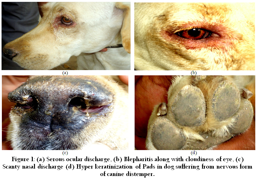
Figure 1: (a)Serous ocular discharge, (b) Blepharitis along with cloudiness of eye, (c) Scanty nasal discharge (d) Hyper keratinization of Pads in dog suffering from nervous form of canine distemper
There is no specific anti–viral drug for this disease so symptomatic treatment was done (http://dogtime.com/distemper–in–dogs–vin.html). The treatment included anti–pyretic (inj. ketoprofen @ 2mg/kg) to reduce the fever, 3rd generation broad spectrum anti–biotic (inj. Ceftriaxone sodium @ 25mg/kg b.w) to avoid secondary bacterial infection. To check diarrhea and dehydration inj. Metronidazole @ 10mg/kg b.w and inf. ringer lactate @ 15ml/kg b.w was given respectively. From 2nd day to onward, with fluid therapy Ketoprofen syrup) @ 2mg/kg b.w once a day, Cephalexin (Keflex syrup) @15mg/kg 2 times a day and gravinat syrup @ 3mg/kg b.w afted feeding was recommended. A multi–vitamin powder (Bendoz powder) was added in feed @ 10g/day to support the animal. Same treatment was repeated for 3rd, 4th and 5th days.Vita A also play an important role for the treatment of canine distemper but mechanism is still unknown (Rodeheffer et al., 2007).
Morbillivirus of family Paramyxoviradae is the causative agent of an infectious disease of carnivore known as Canine distemper can also effect the vaccinated animals. At age of 6–8 month the disease most likely to occur depends on breed of animal (Chappuis, 1995). Not only canine the members of family Mustelidae and Procyonidae also susceptible (Appel, 1987). Most of times the recently weaned pups get this infection because at this stage the maternal immunity that is coming from milk are at its lowest level (Shabbir et al., 2010). Therefore it is recommended to vaccinate the dogs at 3 month of age.
Depending on the age and immune status of host incubation period varies between 1–4 weeks of different strains. About 50% mortality is seen in this disease without clinical signs to severe clinical signs (Appel, 1970, 1987; Krakowka et al., 1980; Moritz et al., 2000). As cold environment favors virus so the disease mostly occur in winter season. High mortality is seen is 63 % in under 1.5 of age in distemper encephalitis cases (Swango, 1989). All body secretions or excretion will contain virus in acute cases. In this case fever, cutaneous rashes, anorexia, diarrhea blisters on the abdominal region ocular and nasal discharge which is serous in nature along with conjunctivitis, blephritis and cloudiness of eye mostlvisible. Gastrointestinal signs become complicated when secondry infection occur. Progressive neurological signs are seen (Greene and Appel, 1998). Myoclonus, ataxia, plegia, and nystagmus are includes in nervous signs (Amude et al., 2007).
CDV main targets are mucous membranes and lymphoid tissue (Appel, 1987). This virus enter in body by air to URT and there in lymph nodes it primarily replicate and causes immunosuppression then spread to epithelium and CNS at about 10 days after transmission (Krakowka et al., 1980). Mainly cause lymphopenia in initial stages. When it reaches to lower respiratory tract (LRT), gastrointestinal tract and CNS cause lesions to be formed on these organs and responsible for appearance of systemic, cutaneous and nervous signs (Greene and Appel, 1998). Out of all CNS inflammation 15% of deaths of dogs caused by encephalomyelitis produce in Canine distemper (Appel and Summers, 1995).CDV produce encephalitic lesions and multifocal demyelination in CNS instead of inflammatory changes cause death of dogs after passing of systemic phase (Beineke et al. 2009).

Presence of distemper myoclonus (Figure 2) and hyper keratinization (Figure 1d) and lymphopenia in blood report confirm the disease as Canine Distemper. Because myoclonus and keratinization of foot pad are considered as the pathognomonic signs of this disease (http://www.2ndchance.info/dogdistemper.htm).
Animal can recover if promote the production of anti–bodies against the virus. But in footpads and lymphatic cells, this virus can persist and cause hyper keratinization (Appel, 1970, 1987; Greene and Appel, 1998) Due to this reason this disease is also called as hard pad disease.
At this time the only thing that we can do to prevent this disease is vaccination. By vaccination at regular intervals will help us a lot to control this disease until the specific treatment is not discover.
ACKNOWLEDGEMENT
The authors would like to acknowledge Dr. Asher Mahfooz Lecturer, (Department of Clinic Medicine and Surgery, University of Agriculture Faisalabad) and Dr. Asim Shahzad Research Associate, (Department of Pathology, University of Agriculture Faisalabad) for their motivation and Support.
REFERENCES
Alex PC and Dhanapalan P (1994). Distemper encephalitis in dogs: incidence, symptomology and electroencephalographic findings. J. Vet. Anim. Sci. 25: 127– 31.
Amude AM, Alfieri AA and Alfieri AF (2007). Clinicopathological findings in dogs with distemper encephalomyelitis presented without characteristic signs of the disease. Res. Vet. Sci. 82: 416– 422.
http://dx.doi.org/10.1016/j.rvsc.2006.08.008
PMid:17084426
Appel MJ (1970). Distemper pathogenesis in dogs. J. Am. Vet. Med. Assoc. 156: 1681– 1684.
PMid:4912306
Appel MJ (1987). Canine distemper virus. In: Horzinek, M.C.M. (Ed.), Virus Infection of Vertebrates, vol. 1. Elsevier Science Publisher B.V., Amsterdam, Oxford, New York, Tokyo, 133– 159.
Appel MJG and Summers BA (1995). Pathogenicity of morbilliviruses for terrestrial carnivores. Vet. Microbiol. 44: 187– 191.
http://dx.doi.org/10.1016/0378-1135(95)00011-X
Beineke A, Puff C, Seehusen F and Baumgartner (2009). Pathogenesis and immunopathology of systemic and nervous canine distemper. Vet. Immunol. Immunopathol. 127: 1– 18.
http://dx.doi.org/10.1016/j.vetimm.2008.09.023
PMid:19019458
Chappuis G (1995). Control of canine distemper. Vet. Microbiol. 44(2/4): 351– 358.
http://dx.doi.org/10.1016/0378-1135(95)00028-9
Elia G, Decaro N, Martella V, Cirone F, Lucente MS, Lorusso E, Trani LD and Buonavoglia C (2006) Detection of canine distemper virus in dogs by real–time RT–PCR. J. Virol. Meth. 136: 171– 176.
http://dx.doi.org/10.1016/j.jviromet.2006.05.004
PMid:16750863
Greene CE and Appel MJ (1998). Canine distemper. In: Infectious diseases of dogs and cats. WB Saunders Company, Philadelphia, London, Toronto, Montreal, Sydney, Tokyo. pp. 9– 22.
Krakowka S, Higgins RJ and Koestner A (1980). Canine distemper virus: review of structural and functional modulations in lymphoid tissue. Am. J. Vet. Res. 41: 284– 292.
PMid:6989302
Lan NT, Yamaguchi R, Unchida K, Sugano S and Tateyama (2005). Growth profiles of recent canine distemper isolates on Vero cells expressing canine signaling lymphocyte activation molecule (SLAM). J. Comp. Pathol. 133: 77– 81.
http://dx.doi.org/10.1016/j.jcpa.2005.01.006
PMid:15899494
Mortiz A, Frisk A and Baumgartner W (2000). The evaluation of diagnostic procedures for the detection of canine distemper virus infection. Eur. J. Comp. Anim. Pract. 10: 37– 47.
Swango LJ (1989). Canine viral diseases. In: Textbook of veterinary internal medicine. 3rd ed. Philadelphia: W.B Saunders Company, Philadelphia, London, Toronto, Montreal, Sydney, Tokyo. pp. 301– 303.
Shabbir MZ, Arfanahmad M, Aliahmed, Muhammad K, Anwar I, 2010. Comparative evaluation of clinical samples from naturally infected dogs for early detection of canine distemper virus. Turk. J. Vet. Anim. Sci. 34(6): 547– 552.
Wright NG, Cornwell AJ, Thompson H and Lauder IM (1974). Canine distemper: current concepts in laboratory and clinical diagnosis. Vet. Rec. 94: 86– 92.
http://dx.doi.org/10.1136/vr.94.5.86
PMid:4593536
Zafar M. S., Khan S. A. and Rabbani A., 1999.Hematological studies and estimation of electrolytes in dogs. P. Vet. J. 19(1): 35– 39.
Rodeheffer C, Messling V, Milot S, Lepine F, Manges AR, Ward BJ (2007). Disease manifestations of canine distemper virus infection in ferrets are modulated by vitamin a status. J.Nutrition 137 (8): 1916– 22
PMid:17634264


