Research Journal for Veterinary Practitioners
Case Report
Res. J. Vet. Pract. 4(2): 34-38
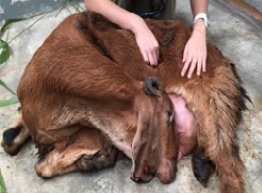
Figure 1
Doe with head tilted to the left flank
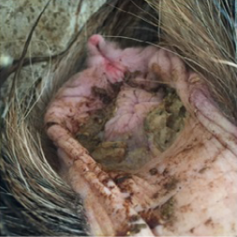
Figure 2
Light brown pasty fecal staining around the perineum region
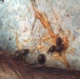
Figure 3
Well-formed feces covered with orange mucoid discharge
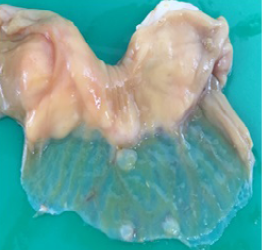
Figure 4
Transparent thinning of the intestinal wall indicative of necrotizing enteritis
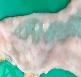
Figure 5
Petechial haemorrhage on the mucosal layer of the intestine
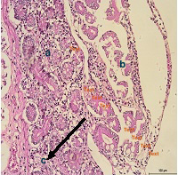
Figure 6
a) Infiltrations of inflammatory cells in the submucosa layer; b) loss of architecture in Crypt of Lieberkuhn; c) necrosis of the Brunners’ gland in the small and large intestine


