Research Journal for Veterinary Practitioners
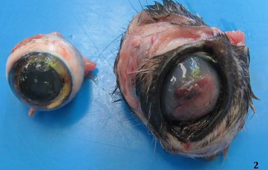
DSH cat presented with intraocular and retrobulbar mass affecting the right eye
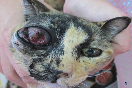
Right intraocular mass with ulcerative keratitis
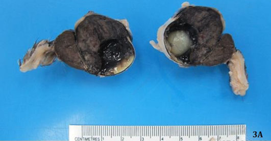
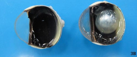
Firm large brown to black retrobulbar and intraocular mass effacing both anterior and posterior chambers of the right eye (Figure 3A) compared to normal left eye (Figure 3B)
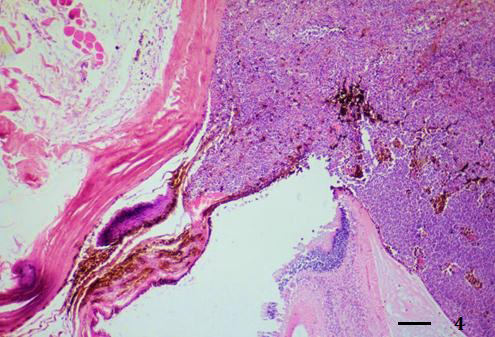
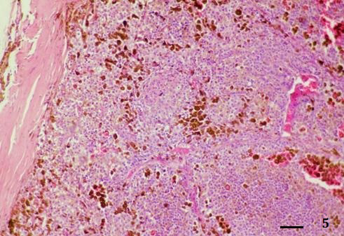
Extensive melanoma occupied iris and posterior chamber (H&E stain, bar = 250 µm)
Diffuse solid nests of tumor cells in posterior chamber (H&E stain, bar = 100 µm)
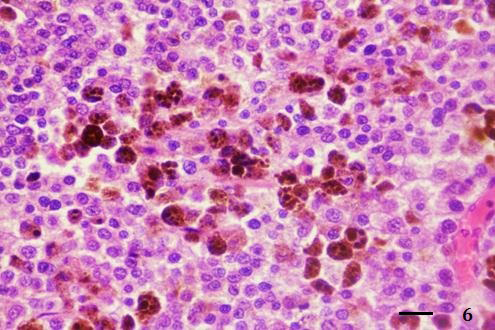
Neoplastic cells had large epithelioid shape and contained intracytoplasmic brown black melanin pigments (H&E stain, bar = 25 µm)
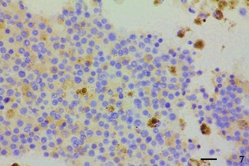
Tumor cells showed intense cytoplasmic Melan-A protein expression (IHC; Envision polymer, counterstained with Mayer’s Hematoxylin, bar = 25 µm)


