Research Journal for Veterinary Practitioners
Case Report
Research Journal for Veterinary Practitioners 2 (6): 108 – 110Surgical Management of a Rare Case of Extensive Obstructive Fibrosarcoma in a Pomeranian Bitch
Rayadurgam Venkata Suresh Kumar1*, Gurram Kamalakar1, Rajapantula Mahesh1, Narpala Sumiran1, Jetty Devaratnam1, A. Anand Kumar
- Department of Veterinary Surgery & Radiology, College of Veterinary Science, Sri Venkateswara Veterinary University, Proddatur, Y S R Kadapa district, Andhra Pradesh, India – 516360
- Department of Veterinary Pathology College of Veterinary Science, Sri Venkateswara Veterinary University, Proddatur, Y S R Kadapa district, Andhra Pradesh, India – 516360.
*Corresponding author:goraya_uaf@yahoo.com
ARTICLE CITATION:
Kumar RVS, Kamalakar G, Mahesh R, Sumiran N, Devaratnam J, Kumar AA(2014). Surgical management of a rare case of extensive obstructive fibrosarcoma in a pomeranian bitch. Res. J. Vet. Pract. 2 (6): 108 – 110.
Received: 2014–06–09, Revised: 2014–07–06, Accepted: 2014–07–08
The electronic version of this article is the complete one and can be found online at
(
http://dx.doi.org/10.14737/journal.rjvp/2014/2.6.108.110
)
which permits unrestricted use, distribution, and reproduction in any medium, provided the original work is properly cited
ABSTRACT
An intact Pomeranian bitch of 12 years age was presented to the hospital with hard perineal swellings, anorexia, dysuria, constipation, tenesmus, distended abdomen. Clinical examination revealed tense abdomen, nodular dermatofibromas, and palpable growths in vagina which were further evaluated by biochemical, haematological, radiological, and ultrasonographic examination. The growths were excised under general anaesthesia by ovario hysterectomy and episiotomy procedures. Histological sections of tumour mass confirmed it as fibrosarcoma.
Tumourous conditions related to reproductive system are common in females compared to male dogs. Most commonly reported tumours of reproductive tract were leiomyosarcomas, fibroleiomyomas, lipomas and squamous cell carcinomas and occur in intact females of around 10 yrs age (Fossum, 2013). But, reports of fibrosarcoma of vagina compared to that of skin and sub cutis in dogs were very low (Mumba et al, 2013). The growths may or may not be peduculated (Al–Kenanny et al, 2013) or grow in concentric way either towards cervix or to exterior (Thacher and Bradley, 1983) and cause obstruction to urethra and rectum extra luminally or intra luminally (white et al, 2008 and Gupta et al, 2014). The signs in such obstruction include dysuria, pollakuria, haematuria, stranguria and constipation. Diagnosis can be made based on clinical signs, radiography (plain or contrast) and ultrasonography. Surgical removal of the tumours along with ovario hysterectomy was indicated in such cases (Fossum, 2013). This paper reports a rare case of extensive fibrosarcoma of uterus, cervix, vestibule and vagina in a bitch and its management by surgical excision of masses along with ovario hysterectomy.
An intact Pomeranian bitch of about 12 years age was presented to the Department of Veterinary Surgery and Radiology, College of Veterinary Science, Proddatur with a history of hard swelling at perineal region around anus and vagina since last one month. The bitch was unable to urinate and defecate freely though the faeces were soft. It was straining severely with tenesmus. It was anorectic since last three days. It was treated by local veterinarian using laxatives and other traditional therapies without any improvement.
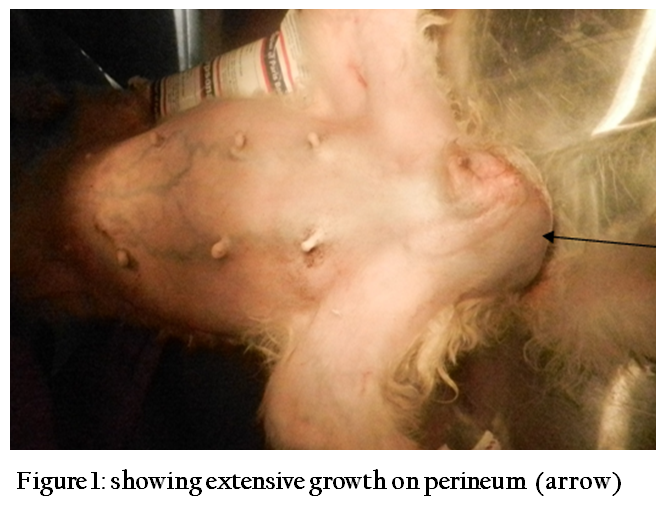
Clinical examination showed that, the dog was dull, dehydrated with tenesmus, stranguria, and pollakuria. Pyrexia (1040F), tachycardia (140/ min), tachypnoea (46/ min), congested conjunctival mucous membrane were observed. The perineal swelling was very hard (Figure 1). Per vaginal palpation revealed no discharges but multiple hard spherical structures in vestibule, vagina up to the level of cervix and appeared to cause extra luminal obstruction of urethra. Per rectal examination revealed soft faeces and no growths in the lumen but growths in the vagina were causing extra luminal obstruction. Few pedunculated growths were observed in the vestibule. The pedunculated growth was sent for biopsy.
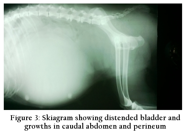
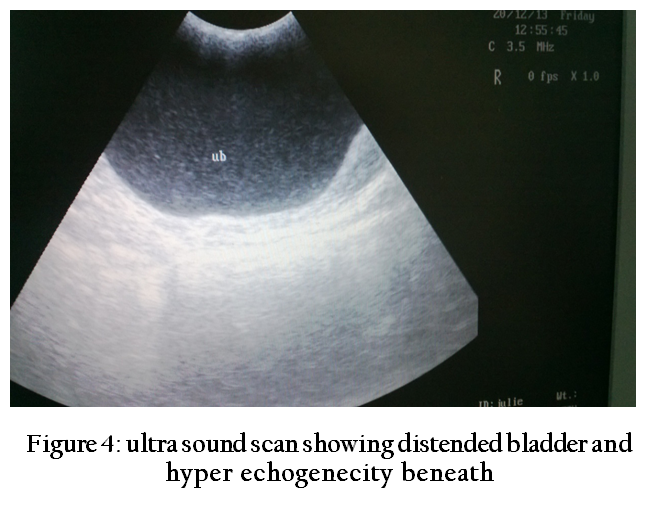
Lateral radiography of abdomen and pelvis revealed distended bladder up to costal arch, and irregular spherical masses in caudal abdomen, pelvic and perineal regions (Figure 3). Thoracic radiographs showed no metastatic lesions. Ultrasonography revealed anechoic distended bladder and irregular hyper echoic masses in abdominal and pelvic regions (Figure 4). The dog was immediately catheterised to relieve urine which was straw coloured, turbid and odd odoured. Urine analysis revealed more of epithelial casts and leucocytes while pH was 6.0. Haemogram showed 9% Hb, normocytic normochromic anaemia and leucocytosis with neutrophilia. Cytological examination revealed malignant cells. Based on the relevant information it was decided to perform exploratory laparotomy and surgical excision of masses to the extent possible as a salvage procedure.
As the haematological and biochemical values are slightly altered, the animal was rejuvenated to fit for surgery by administering inj. Iron dextran, inj. Vitamin AD3E, inj. ceftriaxone and inj. B complex and oral laxatives and appetonics for 10 consecutive days and urethral catheter was kept in situ. After 10 days, the dog was recovered and all the tests were repeated which showed nearly normal values and planned for surgery. The biopsy of the growth confirmed as fibrosarcoma.
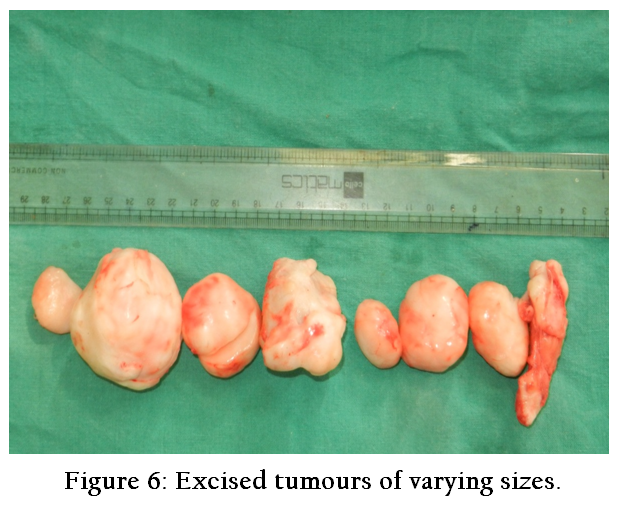
The dog was asceptically prepared for surgery, pre anaesthetised with atropine @ 0.04mg/kg, induced with xylazine and ketamine hydro chloride @ 0.8 & 10 mg/kg respectively and maintained with diazepam @ 0.5 mg/ kg BW. The animal was catheterised through the external urethral orifice. By caudal mid ventral abdominal incision, the uterus with tumour masses were exposed out (Figure 5) and performed ovario hysterectomy by applying modified transfixation ligatures using No. 0 chromic catgut and the abdominal wound was closed in three layers as a routine procedure. The size of the masses ranged between peanut and big lemon size with glistening capsular appearance (Figure 6). Through episiotomy the remaining growths at the level of cervix, vagina and vestibule were excised by applying ligatures with 1/0 plain catgut and episiotomy incision was closed in two layers in simple interrupted manner. Post operatively the animal was infused 250 ml of ringers lactate, 100ml of metronidazole for 3 consecutive days and ceftriaxone 0.5g, meloxicam 1ml and 1.5ml Tribivet were administered and by following dressing povidone ointment for 5 days. The animal recovered without any complications and skin sutures were removed on 10th post operative day.

Histopathology of the mass revealed numerous spindle shaped cells (fibroblasts) with scanty cytoplasm and rounded nuclei. Cells were arranged in interwoven pattern with marked pleomorphism indicating malignancy (Figure 7).

Figure 7: Histopathology showing numerous spindle shaped fibroblasts in interwoven pattern with marked pleomorphism
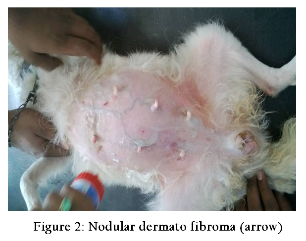
In dogs, tumours of uterus and vagino–vestibular region account for 0.36 and 2.6% respectively (Johnston, 1993). Of these, fibrosarcomas are relatively unusual tumours vagina of bitches of mesenchymal origin ( Neelu and Tiwari, 2009). These growths mostly seen outside and may present concealed in vagina and lead to extra luminal obstruction of urethra and rectum as seen in the case and as reported by Gupta et al (2014). Symptoms of pollakuria, dysuria, stranguria and tenesmus observed like in this case were in conjunction with that of Johnston (1993). These tumours may occur along with other uterine affections like pyometra (Tsioli et al, 2011) but no such observations were in the present case. Radiography and ultrasound examination was used for diagnosis of extent of lesions apart from clinical symptoms, biochemical, haematological and histo pathological findings but, White et al (2008) used contrast vagino – urethrogram and urethral biopsy for diagnosis of obstructive tumours. The growths appeared to be large and present on the whole genital tract, ovario hysterectomy and episiotomy procedures were adopted as reported by Gupta et al (2014). Few small growths were left as they regress in due course after performing ovario hysterectomy (Fossum, 2013). Nodular dermatofibrosis was evident (Figure 2) in the present case may be a metastatic lesion. Lium and Moe (1985) reported nodular dermatofibrosis as a metastatic consequence of renal cystadenocarcinoma in German shepherd dogs.
In this present study a case of extensive fibrosarcoma in a geriatric intact bitch and its surgical management was reported. It was concluded that the urinary and faecal retention by tumours in bitches can be successfully treated with ovario hysterectomy and episiotomy procedures.
REFERENCES
Al–Kenanny ER, Al–Hayani OH, Al–Annaz MTh (2013). Vaginal fibrosarcoma in a bitch: a case report. Iraqi J. Vet. Sci. 27(2): 119 – 121.
Fossum TW (2013). Surgery of the Genital and Reproductive systems, in Small Animal Surgery, In Fossum TW (eds.) 4th Edn, Elsevier Mosby, Philadelphia. pp: 825.
Gupta P, Gupta AK, Kushwaha RB, Dwivedi DK, Sharma A (2014). Urethral and Rectal Obstruction Caused by Vaginal Tumour in a Bitch. Ind. Vet. J. 91(04): 79 – 80.
Johnston SD (1993). Reproductive Systems, in Text Book of Small Animal Surgery, Slatter D (ed.) WB Saunders Co., Philadelphia. pp: 2182.
Lium B, Moe L (1985): Hereditary multifocal renal cystadenocarcinomas and nodular dermatofibrosis in the German shepherd dog: macroscopic and histopathologic changes. Vet. Path. 22(5): 447 – 55.
PMid:4049673
Mumba C, Pandey GS, Chijikwa J, Munjita S (2013): Multiple vaginal fibrosarcoma in a Dog. Ind. Vet. J. 90(2):108 – 109.
Neelu G and Tiwari SK (2009): Study on incidence, histopathological features and surgical managements of neoplasms in canines. Vet. World. 2(10): 393 – 395.
Thacher C, Bradley RL (1983). Vulvar and Vaginal tumours in the dog: A retrospective study. J. Am. Vet. Med. Assoc. 183: 690 – 692
PMid:6629979
Tsioli VG, Gouletsou PG, Loukopoulos P, Zavlaris M, Galatos AD (2011). Uterine leiomyosarcoma and pyometra in a dog. J. Sm. Ani. Pract. 52: 121 – 124.
http://dx.doi.org/10.1111/j.1748-5827.2010.01030.x
PMid:21265853
White RN, Davies JV, Gregory SP (2008). Vagino urethroplasty for treatment of urethral obstruction in the bitch. Vet. Sur. 25: 503 – 510.
http://dx.doi.org/10.1111/j.1532-950X.1996.tb01451.x


