Research Journal for Veterinary Practitioners
Research Article
Research Journal for Veterinary Practitioners 2 (5): 73 – 77Economic Losses in a Commercial Dairy Farm due to the Outbreak of Johne’s Disease in India
Krishna Dutta Rawat1, Sarjeet Chaudhary2, Naveen Kumar1, Saurabh Gupta1, Kundan Kumar Chaubey1, Shoor Vir Singh1*, Kuldeep Dhama3, Rajib Deb4
- Microbiology Laboratory, Animal Health Division, Central Institute for Research on Goats, Makhdoom, PO–Farah, Mathura, India
- District Animal Disease Diagnostic Center, Department of Animal Husbandry, District Alwar, Rajasthan, India
- Division of Pathology, Indian Veterinary Research Institute (IVRI), Izatnagar, Bareilly (U.P.) – 243122, India
- Animal Genetics and Breeding, Project Directorate on Cattle, Meerut – 250 001, Uttar Pradesh, India
*Corresponding author:shoorvir.singh@gmail.com; shoorvir_singh@rediffmail.com
ARTICLE CITATION:
Rawat KD, Chaudhary S, Kumar N, Gupta S, Chaubey KK, Singh SV, Dhama K, Deb R (2014). Economic losses in a commercial dairy farm due to the outbreak of Johne’s disease in India. Res. J. Vet. Pract. 2 (5): 73 – 77.
Received: 2014–04–14, Revised: 2014–05–04, Accepted: 2014–05–06
The electronic version of this article is the complete one and can be found online at
(
http://dx.doi.org/10.14737/journal.rjvp/2.5.73.77
)
which permits unrestricted use, distribution, and reproduction in any medium, provided the original work is properly cited
ABSTRACT
First time in India economic losses due to outbreak (OB) of Johne’s disease (JD) in a Holstein Frisian (H/F) dairy farm were recorded. OB of JD was suspected by clinical signs [loss in body condition, sharp drop in milk yield (30 to 2 litres/day), increase in cases of infertility, mortality etc.] and necropsy findings. JD was confirmed by screening of 30.0% cows by microscopy (68.5%), serum ELISA (92.3%), milk ELISA (60.8%) and blood PCR (35.7%). Losses due to stress culling, mortality and reduced productivity (infertility, stunted growth) were quantified. Growing heifers exhibited weakness, stunting and delayed first heat. Losses due to delayed breeding, reduced fertility and repeat breeding were Rs 1,63,800.0 in 180 days. Losses due to mortality and culling during JD OB were Rs 1,05,000.0 and Rs. 1,67,000.0, respectively. Losses due to reduced milk yield were Rs 54,442.5 /cow/lactation. Total losses at the farm were very high (Rs. 16,87,977.5), since H/F cows being high yielding were at higher risk and suffered with JD outbreak. Low per animal productivity of domestic livestock indicated necessity to initiate JD control programs at the National level.
INTRODUCTION
Mycobacterium avium subspecies paratuberculosis (MAP), the cause of Johne’s disease (JD) is a highly pathogenic mycobacteria affecting dairy cattle and other domestic ruminants globally (Boelaert et al., 2000; Gasteiner et al., 1999; Muskens et al., 2000; Singh et al., 2008; Singh et al., 2013a; Singh et al., 2014a). It is chronic progressive granulomatous infection of high yielding Holstein Friesian (H/F) cows leading to increased culling (Benedictus et al., 1987; Tiwari et al., 2005) decreased milk yield (Benedictus et al., 1987; Tiwari et al., 2007), higher death rates (Kreeger, 1991) and increased susceptibility to other infections (Tiwari et al., 2009). Besides causing major losses in high yielding dairy farms, it has also been associated with number of human diseases. Most common clinical sign in cows is diarrhoea which is continuous along with weight loss. Clinical and asymptomatic cows shed large number of bacilli in feces and milk (Sweeney et al., 1992; Streeter et al., 1995; Slana et al., 2009; Singh et al., 2014a). Colostrum and milk is important source of transmission of MAP to new born calves and human population (Grant, 2003; Ayele et al., 2001; Slana et al., 2008). Calves become infected soon after birth due to higher susceptibility to MAP during first year of life, though clinical symptoms appear at later age (2–6 years). Calves may be infected in-utero from their asymptomatic mother (Buergelt et al., 2006). Effective control of MAP is severely hampered due to lack of indigenous diagnostics and vaccines. It is always challenging to diagnose sub–clinically infected animals using traditional tests (smear examination and intradermal Johnin), because of absence of clinical symptoms to assist interpretation. Use of sensitive fecal culture and serum ELISA tests helped to improve the detection of cases of sub-clinical Johne’s disease. However, development of nucleic acid based diagnostic approaches (IS900 PCR, IS1311 PCR_REA) proved handy in quick confirmation of the disease (Singh et al., 2009). Most frequently used test for JD is based on serum antibody detection using an ELISA platform. Commercially available kits are either costly or poorly sensitive (Singh et al., 2007). Using multiple diagnostic tests, Singh et al. (2014a) reported that disease is endemic in domestic livestock population, therefore effective control program are warranted to stop transmission of disease and reduce the economic losses.
Several studies in western countries reported huge economic losses due to MAP infection (Tiwari et al., 2008, Benedictus et al., 1987; Tiwari et al., 2005). These studies mostly estimated losses based on reduction in milk yield and increased culling and mortality (Tiwari et al., 2005; Tiwari et al., 2007; Kreeger 1991). Though JD is endemic in Indian dairy farms (Singh et al., 2013b; Singh et al., 2014b), however reports on economic losses are non–existent. Present study is maiden attempt to estimate economic losses in a natural outbreak of JD in a high yielding Holstein Frisian dairy farm in Alwar district of Rajasthan in India.
MATERIAL AND METHODS
History of Yadu Dairy Farm
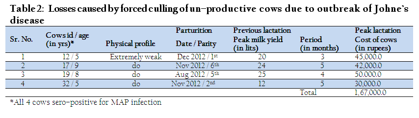
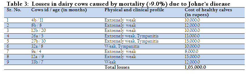
Data from the Yadu commercial dairy farm was collected by personal visits and our first visit to farm was in October, 2013. Dairy farm consisted of Holstein Frisian (H/F) cows was established purely as commercial venture in village Dilawalpur (PO– Shahpur) of Alwar district in Rajasthan by purchasing 10 adults cows and 10 calves from different parts of the country in June 2011. Cows were bred using H/F bulls semen from a semen bank at Bassi in Jaipur. Dairy farm was only source of livelihood for farmer. At the time of JD outbreak the strength of cows in dairy farm was 71 [19 (0–18 m), 11 (18–30 m) and 41 (>30 m)]. Farmer informed that her had to cull four dry cows (5-9 years) that suffered from had poor physical condition, extreme weakness without diarrhoea (except one cow), weight loss, stopped lactating after 3-5 months and did not conceive in this period (table 2). Nine animals (4-20 m age) which died in this period suffered from extreme weakness without exhibiting diarrhea, poor physical condition and tympanitis (table 3). After these losses the attack of JD was suspected by the consulting clinician. On our first visit to dairy farm samples (fecal, blood, serum and milk) of 35 cows (26 adult and 9 calves) were collected and screened for JD, wherein 68.5, 92.3, 60.8 and 35.7% cows were positive for MAP infection in fecal microscopy, serum ELISA, milk ELISA and IS900 blood PCR, respectively (Singh et al., 2014b). Cows exhibiting clinically symptoms of JD were also confirmed by laboratory tests.
Nutritional Status, Breeding, Health and Management of Animals
Cows were maintained under optimum nutrition and intensive management. Cows were provided cultivated green fodder, conserved forage (silage and hay), crop residues and concentrates and were protected from extreme cold and hot weather. Hygienic conditions at the farm were good. Consultancy was provided by qualified doctors from Department of Animal Husbandry, Rajasthan. Cows were regularly vaccinated for Foot and Mouth Disease, Haemorrhagic Septicaemia, de-wormed and dipped against ecto-parasitic infection. Individual cows, as and when falling sick were treated. Cows were artificially inseminated (100.0%) by H/F semen from semen bank at Bassi, Jaipur, Rajasthan.
Economic Profile of Dairy Farm before Infection
Milk Production
Milk yield of cows before and after outbreak of JD was recorded. Healthy cows had no clinical signs and gave good yield of milk at first parity after establishment of dairy farm. However, in next parity lactating cows lost more than 40.0% milk yield in one month period as compared to previous lactation. Loss in milk was more in cows with clinical signs of JD.
Profile of Losses after JD Outbreak
Losses due to JD outbreak were estimated in 31 lactating cows. In previous lactation before JD outbreak, average total milk yield was 407 litres/day and average milk price was (Rs. 25.50/Litre) in Alwar district of (Rajasthan). Parameters such as number of cows, milk yield and cost of milk were treated as fixed values, therefore losses associated with MAP outbreak were estimated precisely across the herd. Effect of MAP sero–prevalence showed a co–relation between MAP infection and milk production (Tiwari et al., 2007) and statistically significant reduction in milk yield was observed after JD outbreak in dairy cows. Average reduction in milk yield was approximately 7 litres/ cow/ day in 305 days of lactation.
Mortality
More than nine percent mortality was recorded in cows and was higher in JD positive cows as compared to JD negative cows. Gross symptoms e.g., thickening of intestinal mucosa and enlargement of lymph nodes were noticed by veterinary practitioner. Cost of cows at the time of death was taken as equivalent to the cost of replacement cow. The carcass value of dead cows was zero. Replacement cost of a healthy cow averaged approximately Rs 40,000.0 per cow in Rajasthan as per the owner of the dairy farm.
Stress Culling
Cows exhibiting clinical symptoms and also positive for MAP infection were removed from the herd to protect others cows from contamination. Losses due to stress culling were 9.8% of the total losses due to outbreak of JD in the farm (table 2 and 5). Average replacement value of the cows was taken as the slaughter value of culled cows though officially cow slaughter is banned in India.
Reproductive Losses
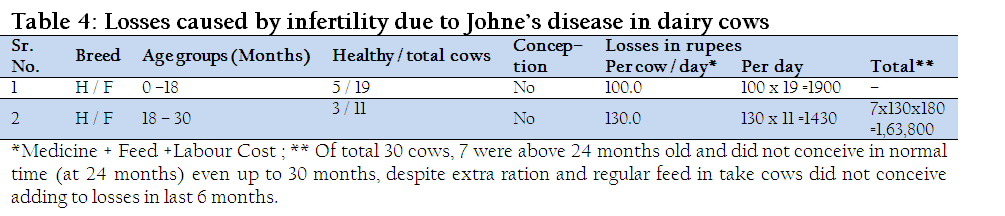
Herd reproductive losses were estimated on the basis of increase in conception period as normal conception age was 24 months in H/F cows. It was observed that 7 cows infected with MAP neither conceived nor showed symptoms of heat up to the age of >30 months. Increased conception period together with herd size, increased calving interval and cost of daily management during increased calving interval estimated the reproductive losses, including extra cost of maintenance of cows, treatment cost and cost in providing improved care and management.
Statistical Analyses
Unpaired t test was used to compare milk yield per month of the infected animals and healthy controls. Value p<0.05 was considered statistically significant.
RESULTS
Outbreak of Johne’s disease was confirmed by screening of 30.0% cows by fecal microscopy (68.5%), indigenous ELISA kit (92.3% in serum and 60.8% in milk) and 35.7% in IS900 blood PCR (Singh et al., 2014b). Irrespective of parity, each cow positive for MAP infection showed significant reduction up to 7 litres/day in milk yield (p<0.05) as compared to healthy cows. Reduction in milk yield was recorded after three weeks of calving.
Average milk production of the dairy herd was (407 litres/day) which reduced to 183 litres/day after the outbreak of JD. A decline of 224 litres /day was recorded in milk production of dairy herd (Table 1) and in terms of value the losses were calculated Rs 54,442.5 /cow/year (305 days of lactation). Economic losses associated with lactating cows were estimated by multiplying the average daily loss of 7 litres of milk / cow in 305 days of lactation with the cost of milk (Rs 25.50 / litre). Cows negative for MAP infection did not show significant (p>0.05) reduction in milk production. Infected cows showed significant reduction (<0.05) in milk yield in the lactation period except first month of lactation as compared to healthy cows (Figure 1).
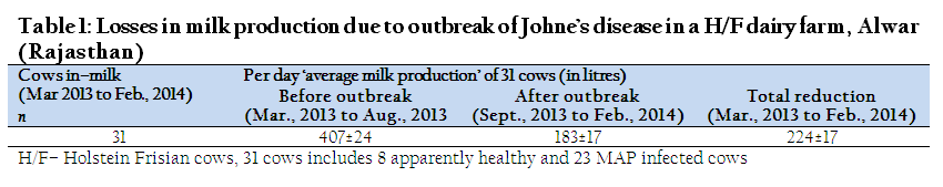
Table 1: Losses in milk production due to outbreak of Johne’s disease in a H/F dairy farm, Alwar (Rajasthan)
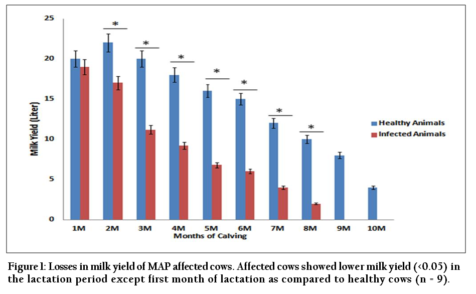
Figure 1: Losses in milk yield of MAP affected cows. Affected cows showed lower milk yield (<0.05) in the lactation period except first month of lactation as compared to healthy cows (n = 9)
Of the total losses (Rs. 16,87,977.5) due to JD outbreak in the dairy farm, losses due to reduction in milk yield were 74.0% (Rs. 12,52,177.5) annually. However, losses due to culling were 9.8% (Rs.1,67,000.0) on yearly basis (Table 2). Similarly annual losses due to mortality and reproductive disorders were 6.2% (Rs. 1,05,000.0) and 9.7 % (Rs. 1,63,800.0), respectively (Table 3, 4 and 5).
DISCUSSION
Percent losses due to mortality and culling in MAP infected cows (sero–positive and endemic) have been documented earlier (Singh et al., 2014), however, economic values of the losses have not been studied in the Indian dairy industry. Clinically early symptoms of Johne’s disease are not visible since the incubation period is long and variable (Tiwari et al., 2005), therefore clinical diagnosis of disease difficult consequently disease continues to spread and gain in severity. This biological complexity leads to increase in MAP infection and economic losses. Benedictus et al. (1987) reported that milk yield of MAP infected animals was affected and milk production was reduced approximately 6% in MAP infected cows at second and third parity. Reduction in milk up to 16.0% has also been reported in cows infected with MAP and confirmed histo–pathologically. Similarly, Abbas et al. (1983) reported 15.0% reduction in milk yield in sub–clinically infected animals as compared MAP negative animals and 4.0% reduction in milk yield was reported in sero–positive animals as compared with sero–negative animals. In contrast, Buergelt et al. (1978) reported no significant reduction in milk yield in culled, asymptomatic, histo–pathologically and fecal–culture positive cows as well as ELISA positive as compared with ELISA negative cows (Johnson et al., 2001).
In the present study marked reduction in milk yield was recorded in those cows that exhibited clinical signs characteristic of Johne’s diseases and were positive for MAP infection in ELISA and further confirmed by PCR as compared to healthy cows. The attack of MAP infection in high yielding cows led to progressive emaciation, decrease in milk production, increase incidence of infertility and mortality rate causing huge economic losses to herd owner. Usually large ruminants infected with JD exhibit symptoms of diarrhoea, which is not treatable, however in the present outbreak of JD, cows suffered from weight loss, weakness and emaciation without diarrhoea except one cow. This was novel feature in large ruminants reported for the first time. Similarly symptoms of continuous diarrhoea, intermittent diarrhoea or weakness without diarrhoea have been recorded by Singh et al., (2014), while monitoring JD in goatherds in last 28 years. MAP infected cows exhibited decreasing trends in milk production and there was significant (p<0.05) drop in milk yield at every month of lactation period, except first month of lactation of the dairy herd (Figure 1). Average reduction in milk production was recorded up to 7 litres/ day/ cow. There was economic loss in milk production to the tune of Rs 54,442.5 per cow annually. Tiwari et al. (2008) reported similar losses ($2992 annually or $49/cow and $409/seropositive cow) in Canadian dairy herds. Another study reported economic losses in milk production and the losses in milk yield increased with increasing parity and in ELISA positive and AFB positive cows (Beaudeau et al., 2007).
Besides losses in milk production, losses were also estimated due forced culling, increased cases of infertility and mortality in cows. Four cows were culled due to attack of JD in one year period in the dairy herd. Average economic losses were calculated as Rs. 1,67,000.0 (9.8%) due to culling of MAP positive cows in a year. Similarly, another study reported economic losses by culling of MAP positive animals (Tiwari et al., 2008, Wilson et al., 1993). Therefore, MAP may be a possible factor in increasing culling rate in Indian dairy herds and may help in culling decision (Beaudeau et al., 2007). In the present study average economic losses due to infertility and reproductive disorders were calculated to the tune of Rs. 1,63,800.0 (9.7%) of the total economic losses to dairy herd including extra medical maintenance cost (Table 5). Other workers also reported economic losses due to infertility. Ifearulundu et al. (2000) reported MAP infected animals appeared progressive emaciated and diarrhoeic coupled with weakness, heat cycle was disturbed which may not be reflected immediately but result in increased infertility. In addition, mortality due to MAP infection also increased economic losses. Mortality risk among dairy herds in MAP positive cows in the present outbreak were recorded at >9.0% in one year. Similarly, Singh et al. (2014a) reported mortality rates of >9.0% in suspected and confirmed cases of JD in Indian goatherds and sheep flocks. In the present study losses due to mortality were calculated as Rs 1,05,000.0 (6.2%) of the total economic losses in the dairy herd due to outbreak of JD. Similarly Tiwari et al. in 2008 reported 3.0% higher mortality rate in MAP infected animals.
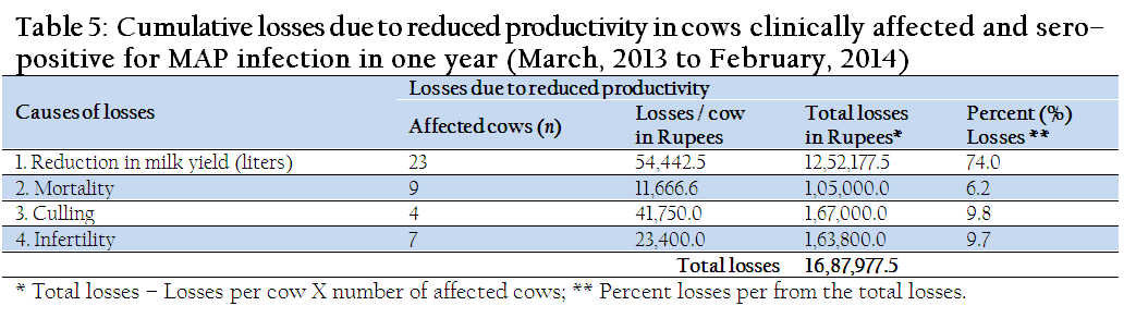
Table 5: Cumulative losses due to reduced productivity in cows clinically affected and sero–positive for MAP infection in one year (March, 2013 to February, 2014)
Present study estimated economic losses due to outbreak of JD in a dairy herd consisting of high yielding H/F cows. Economic losses were calculated on the basis of direct losses (reduction in milk production, increase in cases of infertility and mortality and forced culling of cows). Indirect losses at micro–level if calculated will reveal the actual economic value of the outbreak due to JD and due to endemicity of disease in Indian herds and flocks. The calculated total losses (Rs. 16,87,977.5) were very high therefore monitoring of dairy farms for JD and initiation of JD control programs are immediately required to sustain dairy industry at National level.
CONFLICT OF INTEREST
No conflict of interest to declare.
ACKNOWLEDGEMENTS
Authors are thankful to Council of Scientific and Industrial Research (CSIR, New Delhi) for providing financial assistance and Director, Central Institute for Research on Goats (CIRG), Makhdoom for providing laboratory facilities.
REFERENCES
Abbas B, Riemann HP, Hird DW (1983). Diagnosis of Johne's disease (paratuberculosis) in Northern California cattle and a note of it's economic significance. Calif. Vet. 37: 20–24.
Ayele WY, Machackova M, Pavlik I (2001). The trans¬mission and impact of paratuberculosis infection in domestic and wild ruminants. Vet. Med. 46: 205–224.
Beaudeau F, Belliard M, Joly A, Seegers H (2007). Reduction in milk yield associated with Mycobacterium avium subspecies paratuberculosis (Map) infection in dairy cows. Vet. Res. 38: 625–63.
http://dx.doi.org/10.1051/vetres:2007021
PMid:17565909
Benedictus G, Dijkhuizen AA, Stelwagen J (1987). Economic losses due to paratuberculosis in dairy cattle. Vet. Rec. 121:142–146.
http://dx.doi.org/10.1136/vr.121.7.142
PMid:3660545
Boelaert F, Walravens K, Biront P, Vermeersch JP, Berkvens D, Godfroid J (2001). Prevalence of paratuberculosis (Johne's disease) in the Belgian cattle population. Vet. Microbiol. 77: 269–281.
http://dx.doi.org/10.1016/S0378-1135(00)00312-6
Buergelt CD, Duncan JR (1978). Age and milk production data of cattle culled from a dairy herd with paratuberculosis. J. Am. Vet. Med. Assoc. 173: 478–480
PMid:711590
Buergelt CD, Williams BS, Monif GRG, Pinedo P, Decker JH (2006). Nested polymerase chain reaction and prenatal detection of mycobacterium avium subspecies paratuberculosis (Map) in bovine allantoic fluid and fetuses. Intern. J. Appl. Res. Vet. Med. 4(3): 232–38.
Gasteiner J, Wenzl H, Fuchs K, Jark U, Baumgartner W (1999). Serological cross–sectional study of paratuberculosis in cattle in Austria. Zentralbl Veterinarmed B. 46:457–466.
PMid:10528542
Grant IR (2003). Mycobacterium paratuberculosis and milk. Acta. Vet. Scand. 44(3–4): 261–266.
PMid:15074643
Johnson YJ, Kaneene JB, Gardiner JC, Lloyd JW, Sprecher DJ, Coe PH (2001). The effect of subclinical Mycobacterium paratuberculosis infection on milk production in Michigan dairy cows. J. Dairy. Sci. 84: 2188–2194.
http://dx.doi.org/10.3168/jds.S0022-0302(01)74665-6
Johnson–Ifearulundu YJ, Kaneene JB, Sprecher DJ, Gardiner JC, Lloyd JW (2000).The effect of subclinical Mycobacterium paratuberculosis infection on days open in Michigan, USA, dairy cows. Prev. Vet. Med. 46:171–181.
http://dx.doi.org/10.1016/S0167-5877(00)00145-8
Kreeger JM (1991). Ruminant paratuberculosis– a century of progress and frustration. J. Vet. Diagn. Invest. 3: 373–382.
http://dx.doi.org/10.1177/104063879100300425
PMid:1760477
Muskens J, Barkema HW, Russchen E, Van Maanen K, Schukken YH, Bakker D (2000). Prevalence and regional distribution of paratuberculosis in dairy herds in The Netherlands. Vet. Microbiol. 77: 253–261.
http://dx.doi.org/10.1016/S0378-1135(00)00310-2
Singh AV, Singh SV, Sohal JS, Singh PK (2009). Comparative potential of modified Indigenous, Indigenous and commercial kits for diagnosis of Mycobacterium avium subspecies paratuberculosis in goat and sheep. Indian J. Exp. Bio. 47: 379–382.
PMid:19579805
Singh SV, Gupta S, Chaubey KK, Rawat KD, Kumar N, Sohal JS, Singh S, Tiwari R, Chakraborty S and Dhama K (2014b). Johne's Disease (JD) in a High Yielding Holstein Friesian Cattle Dairy Farm in India. J. Biol. Sci. 14: 195–203.
http://dx.doi.org/10.3923/jbs.2014.195.203
Singh SV, Gupta S, Singh PK, Singh AV, Sohal JS, Kumar N, Kumar A, Chaubey KK and Singh B (2013b). Therapeutic Management of Clinical Bovine Johne's disease Using Goat Based 'Indigenous Vaccine' in Native Hariana Cattle: Case Reports. Adv. Anim. Vet. Sci., 1(1S): 23–28.
Singh SV, Kumar N, Chaubey KK, Gupta S and Rawat KD (2013a). Bio–presence of Mycobacterium avium subspecies paratuberculosis infection in Indian livestock farms. Res. Opin. Anim. Vet. Sci. 3(11): 401–106.
Singh SV, Singh AV, Singh PK, Sohal JS, Singh NP (2007). Evaluation of an indigenous ELISA for diagnosis of Johne's disease and its comparison with commercial kits. Indian J. Microbiol. 47: 251–258.
http://dx.doi.org/10.1007/s12088-007-0046-2
PMid:23100673 PMCid:PMC3450340
Singh SV, Singh AV, Singh R, Sharma S, Shukla N, Misra S, Singh PK, Sohal JS, Kumar H, Patil PK, Misra P, Sandhu KS (2008). Sero–prevalence of Bovine Johne's disease in buffaloes and cattle population of North India using indigenous ELISA kit based on native Mycobacterium avium subspecies paratuberculosis 'Bison type' genotype of goat origin. Comp. Immunol. Microbiol. Infect. Dis. 31: 419–433.
http://dx.doi.org/10.1016/j.cimid.2007.06.002
PMid:17854892
Singh SV, Singh PK, Singh AV, Sohal JS, Kumar N, Chaubey KK, Gupta S, Kumar A, Bhatia AK, Srivastav AK, Dhama K (2014a). Bio–load and bio–type profiles of Mycobacterium avium subspecies paratuberculosis infection in the farm and farmer's herds / flocks of domestic livestock: A 28 years study (1985–2013). Transbound. Emerg. Dis. 61(Suppl. 1): 1–13 (In Press).
Slana I, Liapi M, Moravkova M, Kralova A, Pavlik I (2009). Mycobacterium avium subsp. paratuberculosis in cow bulk tank milk in Cyprus detected by culture and quantitative IS900 and F57 real–time PCR. Prev. Vet. Med. 89: 223–226
http://dx.doi.org/10.1016/j.prevetmed.2009.02.020
PMid:19349086
Slana I, Paolicchi F, Janstova B, Navratilova P, Pavlik I (2008). Detection methods for Mycobacterium avium subsp. paratuberculosis in milk and milk products: a review. Vet. Med. Czech. 53: 283–306
Streeter RN, Hoffsis GF, Bech–Nielsen S, Shulaw WP, Rings DM (1995). Isolation of Mycobacterium paratu¬berculosis from colostrum and milk of subclinically infected cows. Amer. J. Vet. Re¬s. 56: 1322–1324.
Sweeney RW, Whitlock RH, Rosenberger AE (1992). Mycobacterium paratuberculosis cultured from milk and supramammary lymph nodes of infected asymp¬tomatic cows. J. Clin. Microbiol. 30: 166–171.
PMid:1734049 PMCid:PMC265014
Tiwari A, Van Leeuwen JA, Dohoo I R, Keefe G P, Weersink A (2008). Estimate of the direct production losses in Canadian dairy herds with subclinical Mycobacterium avium subspecies paratuberculosis infection. Can. Vet. J. 49: 569–576.
PMid:18624066 PMCid:PMC2387260
Tiwari A, VanLeeuwen JA, Dohoo IR, Keefe GP, Haddad JP, Scott HM, Whiting T (2009). Management risk factors associated with Mycobacterium avium subspecies paratuberculosis in Canadian dairy herds. Prev. Vet. Med. 88: 32–41
http://dx.doi.org/10.1016/j.prevetmed.2008.06.019
PMid:18692926
Tiwari A, VanLeeuwen JA, Dohoo IR, Stryhn H, Keefe GP, Haddad JP (2005). Effects of seropositivity for bovine leukemia virus, bovine viral diarrhoea virus, Mycobacterium avium subspecies paratuberculosis, and Neospora caninum on culling in dairy cattle in four Canadian provinces. Vet. Microbiol. 109: 147–158.
http://dx.doi.org/10.1016/j.vetmic.2005.05.011
PMid:15970402
Tiwari A, VanLeeuwen JA, Dohoo IR, Keefe GP, Haddad JP, Tremblay R, Scott HM, Whiting T (2007). Production effects of seropositivity of pathogens causing bovine leucosis, bovine viral diarrhea, paratuberculosis, and neosporosis. J. Dairy. Sci. 90: 659–669.
http://dx.doi.org/10.3168/jds.S0022-0302(07)71548-5
Whittington RJ, Marsh IB, Whitlock RH (2001). Typing of IS1311 polymorphisms confirms that bison (Bison bison) with paratuberculosis in Montana are infected with strain of Mycobacterium avium subspecies paratuberculosis distinct from that occurring in cattle and other domestic livestock. Mole. Cell. Probes. 15: 139–45.
http://dx.doi.org/10.1006/mcpr.2001.0346
PMid:11352594
Wilson DJ, Rossiter C, Han HR, Sears PM (1993). Association of Mycobacterium paratuberculosis infection with reduced mastitis, but with decreased milk production and increased cull rate in clinically normal dairy cows. Am. J. Vet. Res. 54: 1851–1857.
PMid:8291762


