Research Journal for Veterinary Practitioners
Short Communication
Research Journal for Veterinary Practitioners 2 (4): 55 – 57Topographic Anatomy of Visceral Organs of a Spotted Deer (Axis axis)
Subrata Kumar Shil1*, Salima Ferdows2, Bibek Chandra Sutradhar3, Bhajan Chandra Das3
- Department of Anatomy and Histology, Chittagong Veterinary and Animal Sciences University, Khulshi, Chittagong–4225, Bangladesh
- Department of Pathology and Parasitology, Chittagong Veterinary and Animal Sciences University, Khulshi, Chittagong–4225, Bangladesh
- Department of Medicine and Surgery, Chittagong Veterinary and Animal Sciences University, Khulshi, Chittagong–4225, Bangladesh
*Corresponding author:skshilvet@yahoo.com
ARTICLE CITATION:
Shil SK, Ferdows S, Sutradhar BC, Das BC (2014). Topographic anatomy of visceral organs of a spotted deer (axis axis). Res. J. Vet. Pract. 2 (4): 55 – 57.
Received: 2014–02–25, Revised: 2014–03–19, Accepted: 2014–03–21
The electronic version of this article is the complete one and can be found online at
(
http://dx.doi.org/10.14737/journal.rjvp/2014/2.4.55.57
)
which permits unrestricted use, distribution, and reproduction in any medium, provided the original work is properly cited
ABSTRACT
One carcass of male spotted deer (Axis axis) was examined to visualize the topographic position of different visceral organs of thoracic and abdominal cavity and to compare these with other ruminants. Examination revealed that apical lobe of left and right lung was extended up to 2nd intercostal space and 2nd rib respectively. Base of the heart was extended from mid of 3rd intercostal space to the level of cranial border of 6th rib and apex was at the level of 6th chondrosternal junction. Laterally diaphragm was attached at the mid of 11th rib to 13th rib. Liver was extended from the lumbocostal angle to the level of 7th costochondral junction and ventral border was at the level of each costochondral junction of last rib to 7th rib of right side. Gall bladder was absent. Spleen was extended from the proximal part of 11th intercostal space to the distal third of 8th intercostal space. Right kidney was located below the level of 1st, 2nd and 3rd lumbar transverse processes whereas left was below the level of 2nd to the 4th lumbar vertebrae. These findings may guide our zoo veterinarians to make any decision on surgical corrections if needed in spotted deer.
Topographic anatomy is the study of anatomy based on regions or divisions of the body, emphasizing the relations between various structures in that region. Topographic position facilitates to locate and examine deeper organs. Spotted deer, the wild ruminants under the Cervidae family, are popularly found in zoos throughout the world. They are one of the most important wild artiodactylid groups in the world (Fowler and Boever, 1986).
Recently various species of cervids have been farmed for by products, such as antler velvet for oriental medicine (Fowler and Boever, 1986). In spite of being a true ruminant they have no gall bladder except in the mask deer (Fowler and Boever, 1986) and they can run fast as like the animal of simple stomach. Topographic anatomy is important for a zoo veterinarian for their clinical examination, treatment and surgical intervention in some cases. Therefore, the present study was undertaken to reveal the topographic anatomy of visceral organs of the spotted deer and to compare with the organs of domestic ruminant species.
An adult male Spotted Deer (Axis axis) was brought in Sahedul Alam Quadery teaching veterinary hospital, Chittagong Veterinary and Animal Sciences University, Khulshi, Chittagong, Bangladesh for surgical treatment. History revealed that it was a wild spotted deer and during seeking of feedstuffs, local inhabitants of Raozan area, Chittagong, hit it by bamboo stick with an intention to slaughter it for consumption. Upazila Livestock Officer of Raozan office, Chittagong have rescued and hospitalized it. Thorough examination found the deer injured in different body regions with severe multiple left metatarsal fractures. It was treated well but died after four days of treatment. Before post mortem examination species name, tentative age, body weight, body length, external features were recorded in hospital case record sheet. It was then allowed to anatomical study followed by post mortem examination in Anatomy laboratory of the same university. Keeping in dorsal recumbancy, a longitudinal incision was made at the ventral midline from pharynx to the pelvic inlet to remove the skin. Then it was kept in lateral recumbancy and the limbs, muscles were removed carefully without any damage the rib cage. Then the intercostal muscles were removed to easy visualize the topographic position of the organs of the thoracic cavity. Subsequently the diaphragm and muscles of lateral abdominal wall was removed to identify the relative position of visceral organs of abdominal cavity sequentially without any damage or distortion. Different views were examined to describe the organs. Length of the respective organ was measured by a calibrated scale placing on along the long axis of the organ. To describe the position of some organs, red threads were used to encircle the area.
Left and right lung were 25 cm and 29 cm long respectively. Apical lobe of left lung was extended up to 2nd intercostal space whereas apical lobe of right lung was extended upto the level of 2nd rib. Basal lobe of both the lung was curved extending from the base of 11th rib to the level of 7th costochondral junction.
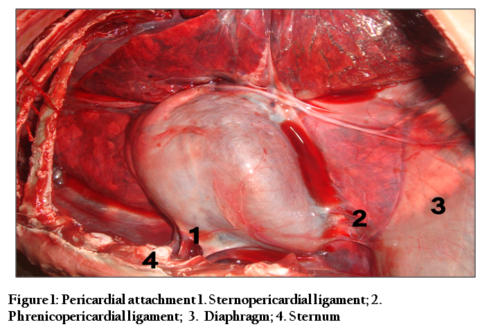
Base of the heart was extended from mid of 3rd intercostal space to the level of cranial border of 6th rib and apex was at the level of 6th chondrosternal junction. Pericardium was attached with both the sternum by two divergent sternopericardial ligaments and diaphragm by phrenopericardial ligament (Figure 1). Though the location of heart is similar to cattle but differ in pericardial attachment. In cattle, the pericardium is attached by sternopericardiac ligaments to the sternum only (Budras et al., 2003). On the other hand, in dog the pericardium is attached with sternum by sternopericardial ligament and with diaphragm by the phrenicopericardial ligament (Evans, 2010). In spite of being a ruminant, the deer showed some similarities with dog in this context. This dual attachment of pericardium may support their heart strongly during their anti-predatory racing in adverse condition.
Dome shaped diaphragm was attached ventrally with xiphoid process at the level of 7th rib. Lateral attachment of both sides was with the 10th rib at the level of costochondral junction and from 11th rib to 13th rib it was attached at mid point. Dorsal attachment was with lumbar vertebrae by two tendinous crura (right and left) at the 2nd to 6th lumbar vertebrae (Figure 2). The right and left crura, the right crus was attached with the first four lumbar vertebrae, the left crus was attached with the ventral the first and second lumbar vertebrae. But in domestic ruminant right crus is attached with upto 4th lumbar vertebrae (Getty, 1975). This tight attachment may provide extra strength to their diaphragm during racing.

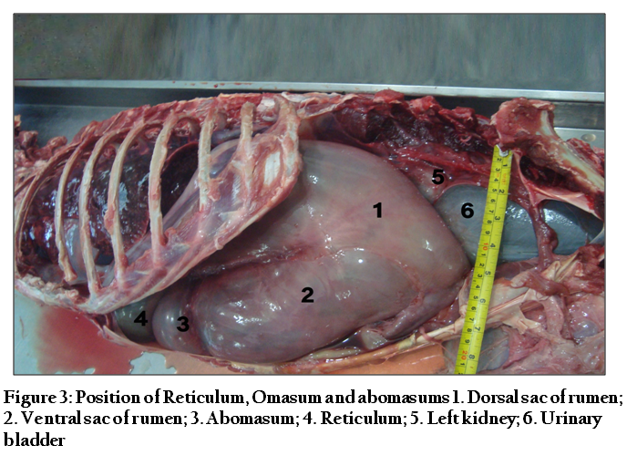
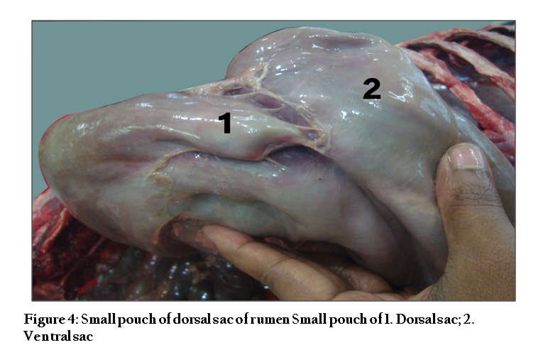
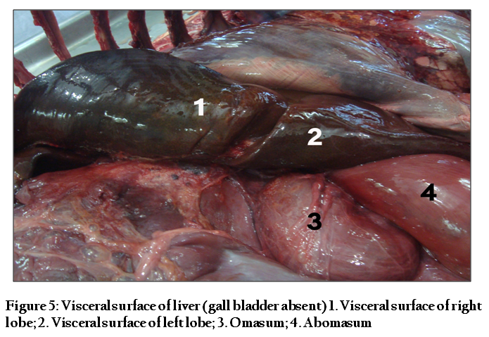
Rumen was extended from 7th intercostal space to the level of coxal tuber, occupied most of the left portion of the abdominal cavity (Figure 3). It was somewhat smaller in compared to its body size. However, a small pouch was noticed on its ventral surface dissimilar to any other ruminants (Figure 4). Reticulum, the most cranial part, was located between the level of 6th and 8th ribs. The greater part of it lied on the left of the median plane. It was somewhat piriform, but was compressed craniocaudally. The diaphragmatic surface lied against the diaphragm and liver. Bean shaped omasum was situated at the right side of the median plane from the level of 7th to 11th rib of right side (Figure 5). Abomasum, the glandular part, was on the abdominal floor dorsal to the xiphoid process. Here, the location and position of various chambers of stomach show similarities with cattle (Habel et al., 1975) and sheep (May et al., 1970). But the shape of omasum varies considerably, as in ox the omasum is ellipsoidal in form (Habel et al., 1975) and in sheep it is ovoid (May et al., 1970). Obliquely and cranioventrally directed liver was 26 cm long measured from caudate lobe to cranial border of left lobe whereas maximum width was 15 cm. Its dorsal border was extended from the lumbocostal angle to the level of 7th costochondral junction and ventral border was at the level of each costochondral junction of last rib to 7th rib of right side. In cattle its ventral border extends upto 6th intercostal space (Habel, 1975). However, this organ shows similarities in position with that of the cattle. On the other hand, gall bladder was absent on the visceral surface of liver (Fig. 5) which completely differ from the ruminant species. Among domestic animals only horse has no gall bladder (Sisson, 1975a).
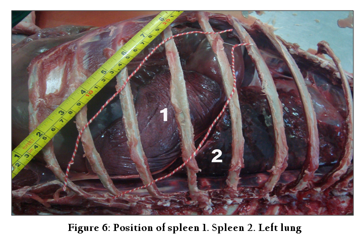
Spleen was large, elliptical in outline placed on the dorsal surface of the rumen obliquely in cranioventral direction. It was extended from the proximal part of 11th intercostal space to the distal third of 8th intercostals space (Figure 6). But, in cattle, the upper extremity of spleen lies under the dorsal end of the 12th and 13th rib of left side and the lower extremity is opposite to the 8th or 9th rib (Sisson, 1975b). On the other hand, in sheep and goat it is approximately triangular in shape that extends obliquely from the vertebral end of the last rib to about the middle of the 10th intercostal space or 11th rib (Sisson, 1975b). Thus, location and shape of spleen of deer varies significantly with that of small ruminant whereas it shows dissimilarities only in position with cattle.
Right kidney was located below the level of 1st, 2nd and 3rd lumbar transverse processes. But the left kidney was located dorsal to the rumen below the sublumbar muscles extending from the level of 2nd lumbar to the 4th lumbar vertebrae, suspended in a fold of mesentery. These findings disagree with Sisson, (1975c) as in cattle, the right kidney lies beneath the proximal part of 13th rib to the 3rd lumbar transverse process and left kidney lies beneath the 3rd to 5th lumbar vertebrae. This study also revealed the smooth surfaced elongated bean shaped kidney similar to kidneys of sheep and goat and dissimilar to kidneys of cattle (Sisson, 1975c). These findings support the Halder et al., (2002).
In spite of being a ruminant, the spotted deer showed some differences in anatomical position, location and shape of some visceral organs in compared with domestic ruminants, notably attachment of pericardium with sternum and diaphragm, absence of gall bladder, comparatively smaller rumen, bean shaped omasum, smooth surfaced kidney, tight attachment of diaphragm with lumbar vertebra etc.
ACKNOWLEDGEMENT
The authors are grateful to the DR. Abdul Mannan, Upazila Livestock Officer, Raojan, Chittagong, Bangladesh for his kind co–operation during the treatment process and subsequent postmortem examinations.
CONFLICT OF INTEREST
No conflict of interest
REFERENCES
Budras D and Habel E (2003). Bovine Anatomy, Schlütersche GmbH & Co, Germany. 69–74
Evans HE and Lahunta A (2010). Guide to the dissection of the Dog. 7th ed., W.B.Saunders Co., Missouri, 115
Fowler ME and Boever WJ (1986). Cervidae. In: Zoo and wild animal medicine. 2nd ed, W. B. Saunders Company, London, 981
Getty R (1975). Ruminant Myology. In: R. Getty (ed.), Sisson and Grossman's The Anatomy of The Domestic Animals, 5th ed., W.B. Saunders Company, USA. Volume 1: 820.
Habel RE (1975). Ruminant Digestive System. In: R. Getty (ed.), Sisson and Grossman's The Anatomy of The Domestic Animals, 5th ed., W.B. Saunders Company, USA. Volume 1: 861–915.
Halder D, Roy M, Mahata TK and Bhattacharjya MK (2002). Gross anatomical study on kidney of spotted Deer (Cervus axis). Journal of Interacademicia, 6: 656–659
May NDS (1970). Anatomy of the sheep. University of Queensland Press, Brisbane.
Sisson S (1975a). Equine Digestive System. In: R. Getty (ed.), Sisson and Grossman's The Anatomy of The Domestic Animals, 5th ed., W.B. Saunders Company, USA. Volume 1: 454–497.
Sisson S (1975b). Spleen. In: Getty R. (ed.), Sisson and Grossman's The Anatomy of The Domestic Animals, 5th ed., W.B. Saunders Company, USA. Volume 1: 1063
Sisson S (1975c). Ruminant Urogenital System. In: R. Getty (ed.), Sisson and Grossman's The Anatomy of The Domestic Animals, 5th ed., W.B. Saunders Company, Philadelphia USA. Volume 1: 937–939.


