Research Journal for Veterinary Practitioners
Research Article
Research Journal for Veterinary Practitioners 2 (3): 44 – 48Prevalence of Gastrointestinal Nematodes in Horses of Jabalpur Region
Khelendra Singh Yadav, Pramod Chandra Shukla, Devendra Kumar Gupta, Aditya Mishra*
-
College of Veterinary Science and Animal Husbandry, Nanaji Deshmukh Veterinary Science University, Jabalpur– 482001 (M.P.)
*Corresponding author:amishra5@yahoo.co.in
ARTICLE CITATION:
Yadav KS, Shukla PC, Gupta DK and Mishra A (2014). Prevalence of gastrointestinal nematodes in horses of Jabalpur region. Res. J. Vet. Pract. 2 (3): 44 – 48.
Received: 2013–09–30, Revised: 2014–02–02, Accepted: 2014–02–13
The electronic version of this article is the complete one and can be found online at
(
http://dx.doi.org/10.14737/journal.rjvp/2014/2.3.44.48
)
which permits unrestricted use, distribution, and reproduction in any medium, provided the original work is properly cited
ABSTRACT
The present study showed an overall prevalence of gastrointestinal nematodes in horses was found as 59.25% (80/135). On the basis of mean egg per gram (EPG), the higher prevalence was found in unorganized sector i.e., 65.45% in comparison of organized sector i.e. well managed farms (32%). Species of nematodes identified in the study included single infestation like, Strongyle (25%), Parascaris equorum (18.75%) followed by mixed infestation like Strongyles and Parascaris equorum (47.50%), Strongyles (species identification not done), Strongyloides and Parascaris equorum (5.0%) and Strongyle and Strongyloides (3.75%). The results of the present investigation revealed a relatively higher prevalence of mixed infestation in the horses. The prevalence of gastrointestinal nematodes in horses was maximum in the age group of 1–6 years (69.09%) followed by 6–12 years (62.96) and above 12–18 years (34.78%) respectively. The highest mean EPG was found in 12–18 years (1229.2) followed by 1–6 years (1079.50) and 6–12 years (887.29). The prevalence of gastrointestinal nematodes in horses was higher in females (60.97) in comparison to males (58.51). The highest mean EPG was found in females (1162.51) in comparison of males (942.24).
INTRODUCTION
Gastrointestinal nematodes are becoming increasingly resistant to the anthelmintics used to control them. The group of equine strongyle, Strongyloides and Parascaris equorum nematodes is very diverse and consists of about 60 described species (Stoltenow and Purdy, 2006). Climate variation, pasture and stable management, anthelmintic treatment and nutritional status of horses are the major epidemiological features which have been recognized for nematode infections. The cost of routine vermifuge applications on herds, the problem of residues in animal products and the environment have prompted for research on the anthelmintic activity of plant extracts. The anthelmintic efficacy of the aqueous extracts of neem (Azadirachta indica) leaf, stem, root and barks against the hatching of eggs and the survival of larvae of nematode parasites of small ruminants have been studied. In the present investigation work was primarily designed to study the prevalence of gastrointestinal nematodes in horses in and around Jabalpur, M.P., India.
MATERIALS AND METHODS
The prevalence of gastrointestinal nematodes of horses was studied in nine private and two government sector farms located in and around Jabalpur, M.P., India. In the present study, a total of 135 horses were screened for finding the prevalence and included about 20 horses every month for a period of six months to study the month wise variation of nematodes in horses. For the proposed work, 20–30 g of faecal sample (freshly voided) was collected from each animal. These samples were packed in small polythene bags and ligated with rubber bands and after proper labeling these polythene bags were brought to laboratory for further processing and examination. The age, sex, history of deworming, feeding habbits, managemental and health status of the animals were also recorded.
Faecal Examination by Qualitative Method
Macroscopic Examination
Faecal samples collected from the horses were placed in a clean glass petridish and examined for the presence of any adult nematodes. It was also examined to determine the consistency and any type of abnormality of the faecal sample.
Microscopic Examination
Direct smear technique: A small quantity of crushed faecal sample (about 3 g) was mixed with 10 ml of water in a beaker, stirring was done continuously and then a drop of this solution was put on a clean glass slide, which was covered with a cover slip. Examination was done under low power of compound microscope (10 X) for the presence of eggs or larvae of any parasite in the faecal sample.
Flotation Method
Nematode and cestode eggs were detected by modified Sheather’s sugar floatation technique (Sloss and Kemp, 1997). Three gram of faecal sample was mixed in 30 ml of water and strained through a tea strainer. After sieving 1–2 ml of suspension was taken in a 15 ml centrifuge tube, then sheather’s solution was added up to 3\4 of the tube. The tube was agitated carefully by putting in between the thumb and forefinger, more sheather’s solution was added up to the brim of the tube making a convex layer. A circular cover slip was placed over the tube and centrifuged at 1500 rpm for 3–4 min. After centrifugation, cover slip was lifted vertically and kept on a glass slide and then examined for the presence of eggs.
Faecal Examination by Quantitative Method
Modified Mc Master Technique:To assess the severity of infection in the positive animal eggs per gram of faeces (EPG) was counted for each positive sample by Modified McMaster Technique (Soulsby, 1982).
RESULTS
Prevalence of Gastrointestinal Nematode
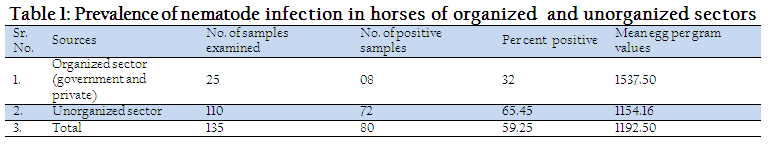
The overall prevalence of gastrointestinal nematodes in horses in and around Jabalpur in Madhaya Pradesh, India, was recorded and revealed that out of 135 horses 80 were positive, hence overall prevalence of gastrointestinal nematodes recorded in the present study was 59.25% (Table No.1).
During the present study, a total of 135 samples were examined involving both (organized and unorganized) sectors i.e. 25 samples from organized sectors and 110 samples from unorganized sectors indicating the relative prevalence of 32% and 65.45% in organized and unorganized sectors, respectively (Table No.2)
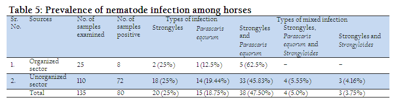
On the other hand the mixed infection of Strongyles and Parascaris equorum was found to be 62.5% and 45.83% in organised and unorganized sector, respectively. Whereas the mixed infection of Strongyle, Parascaris equorum and Strongyloides (species identification not done) was 5.55% and that of Strongyle and Strongyloides was observed to be 4.16% in unorganized sectors. (Table No.5).
Age
The prevalence of gastrointestinal nematodes in horses was higher in the age group 1–6 years (69.09%) followed by 6–12 years (62.96) and above 12–18 years (34.78%), respectively (Table No.2).
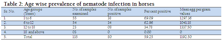
The gastrointestinal nematodes infection was most common in the age group between 1–6 years and 6–12 years. The highest mean EPG was found in 12–18 years (1337.50) followed by 1–6 years (1297.36) and 6–12 years (1041.18). Data on age related prevalence indicated no difference (p = 0.10) among various age groups. A higher proportion of young animals (≤ 10 years of age) were found positive for strongylosis as compared with older horses (≥ 11 years of age).
Sex
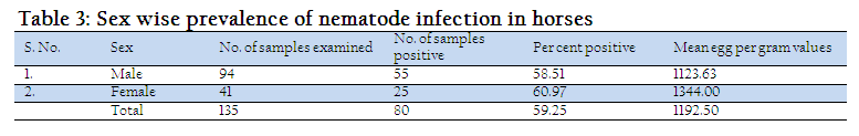
The prevalence of gastrointestinal nematodes in horses was found to be higher in female horses (60.97) in comparison to male horses (58.51). The highest mean EPG values were found in female horses (1344.00) in comparison to male horses (1123.63). (Table No.3)
Season
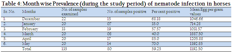
The high prevalence rate of nematodes infection in horses, in the present study was recorded in April (85%) and May (70%) months which might be due to the fact being the peak and favorable months for these infections. (Table No.4)
Feeding Habits and Management
The prevalence was higher in horses where free range system was in practice, whereas prevalence was found quite low where stall feeding was practiced. Among the predisposing factors of internal parasites infection are climates, nutritional deficiency, grazing habits, immunological status, pasture management, presence of intermediate host and vector and the number of infective larvae and eggs in the environment, moreover, damages inflicted to the health and productivity includes loss in body weight, poor reproductive performance, digestive disturbance and emaciation for longer period.
DISCUSSION
Prevalence of Gastrointestinal Nematode
The overall prevalence of gastrointestinal nematodes observed in the present study have also been reported by Khajuria et al., (2004) and Pandit et al., (2008) in Jammu and Kashmir, Katoch et al., (2006) in Haryana and (Sengupta and Yadav, 1998) in Himachal Pradesh in their studies. Further, Singh et al. (2012) found 75.73% which is higher than the results of the present study. They have found that out of total 532 faecal samples examined, 394 were positive with a prevalence of 74.06 per cent which differs from findings of Matto et al. (2013) who have reported as 38.79%. The reason for this increase may be attributed to the large number of animals taken under study by them.
The relative prevalence of different gastrointestinal nematodes in the present study did not reveal any marked difference between these sectors as also studied by Sharma et al. (2011). The observations regarding the feeding practices revealed higher prevalence in horses where free range system was in practice, rather than the animals kept under stall feeding system. Similar observations have also been reported by (Sharma, 2005).
On the other hand the mixed infection of Strongyles and Parascaris equorum, and the mixed infection of Strongyle, Parascaris equorum and Strongyloides observed in the present findings differ from Wannas et al. (2012) who have reported, amongst the parasites detected in horses, the prevalence of Strongylidae, Parascaris equorum, Strongyloides westri and Trichostrongylus axei were 50%, 40.90%, 22.72%, and 25%, respectively and were higher than the findings of the present study. The probable reason for the findings may be that the samples included have been collected from both the sectors during the present investigation.
Studies on prevalence of horse helminths in different parts of world have indicated varied prevalence under different management and parasite control systems (Chaudhry et al., 1991; Montenaro et al., 2002; Champman et al., 2002; Boxell et al., 2004 and Capewell et al., 2005). The managerial practices also reported to have major significance. In the present study, it has been observed that managemental practices viz deworming, feeding, sanitation, housing, hygiene, health coverage and medication wherever, adopted in routine and judiciously the animals found to be comparatively less prone to infection than the other animals kept in unorganized sectors which were lacking in this aspects.
Age
The prevalence of gastrointestinal nematodes in different age groups of horses obtained in the present study is similar to the results obtained by Saeed et al. (2010) and they reported as 75%, 70%, and 42% with average EPG 583, 352, and 354 in the age groups below 3 years, below 10 years and above 11 years, respectively. However, the observation of Ferdowsi et al. (2011) are not in conformity with our findings as during their study the infection rate was found to be 11.34% in stallion and 2.7% in mares. The age wise infection rate was reported to be 6.85%, 7.14% and 10.71% respectively in up to 3 years old, 3–10 years old and over 10 years old horses.
Similarly no effect of age for the strongyle infections could be detected in other studies (Francisco et al., 2009). In one study, small strongyle infections were more common in young horses as compared with mature animals (Bucknell and Gasser, 1995). Severity of infection as determined by EPG has shown a significantly higher average egg excretion by young horses (≤ 3 year old horses) as compared with older horses in other parts of the world (Lind et al., 1999). Higher infection rates and more severe infections indicate a lack of immunity in younger population (Urquhart et al., 1996). The higher prevalence of nematodes in horses of 1–6 years and 6–12 years age observed in the present study might be due to the greater tendency of these horses to use permanent pastures, a high density of horses per unit of pasture and a great turnover rate of incoming and outgoing horses as they were being regularly used for draught purposes.
Sex
The prevalence and mean EPG values of gastrointestinal nematodes in horses were found to be higher in female horses in comparison to male horses. Similar findings have also been reported by (Singh et al., 2012) who have found higher prevalence in female equines (75.73%) as compared to males (72.30%). These observations are in agreement with those reported by (Love and Duncan, 1992; Smith, 2002 and Singh et al., 2012) though the stratification of age has not revealed different in males but statistical difference in various age groups in female was found by Saeed et al., 2010.
Season
The high prevalence rate of nematodes infection in horses, in the present study, recorded in April (85%) and May (70%) months which might be due to the fact being the peak and favorable months for these infections. These findings tallied with the results obtained by (Singh et al., 2012) they have reported the higher intensity of infection in monsoon (79.35%), summer (69.23%) while it was lowest in winter (59.70%). Matto et al. (2013) found the overall prevalence of helminthes as 20.63% with higher rate of occurrence in monsoon (31.29%) followed by winter (20.40%) and summer (14.23%) (Strongyloides (13.19%) and Parascaris equorum (0.23%). On the other hand, Sengupta and Yadav, 1998 have mentioned high rainfall, optimum temperature and high relative humidity favour the development and survival of the infective larvae in and around the region of study.
Relatively higher infection rates and significantly higher egg shedding (p≤0.001) were observed in the present study during spring and summer and findings were consistent with the results of Herd, 1990 who observe that month wise prevalence indicated higher Strongyle infections and more intensity in the month of May as compared with other months. The higher prevalence of helminthes parasite infection during summer and rainy season might be due to high temperature and moisture content which favors the growth and development of larvae on pasture resulting in increased contact between the host and parasite. Season of the sampling does not affect prevalence of strongyles infection as reported by Montenaro et al., 2002; Saeed et al., 2010 and Ferdowsi et al., 2011.
Feeding Habits and Management
The prevalence was higher in horses where free range system was in practice, whereas prevalence was found quite low where stall feeding was practiced. Among the predisposing factors of internal parasites infection are climates, nutritional deficiency, grazing habits, immunological status, pasture management, presence of intermediate host and vector and the number of infective larvae and eggs in the environment, moreover, damages inflicted to the health and productivity includes loss in body weight, poor reproductive performance, digestive disturbance and emaciation for longer period (Radostits et al., 1994). A thorough understanding of the epidemiology of horse helminths under local management and climatic conditions will help in devising effective and economically viable parasite control programs (Saeed et al., 2010).
Climatic variations, pasture and stable management, anthelmintic treatment and nutritional status of horses are the major epidemiological and management features which have been recognized. It has also been found that the sex of the host, parasitological differences in horses seem to be associated with specific management e.g. stable vs. pasture the stable–infection is also possible with contaminated hay or bedding (Smith, 2002).
REFERENCES
Boxell AS, Gibson KT, Hobbs RP and Thompson RCA (2004). Occurrence of gastrointestinal parasites in horses in metropolitan Perth, Western Australia. Aust. Vet. J. 82: 91–95.
http://dx.doi.org/10.1111/j.1751-0813.2004.tb14653.x
PMid:15088968
Bucknell DG and Gasser RB (1995). The prevalence and epidemiology of gastrointestinal parasite of horses in victoria, Australia. Int. J. Parasitol. 25 : 711–724.
http://dx.doi.org/10.1016/0020-7519(94)00214-9
Capewell LG, Hunt D, Guerrero J, Newcomb K and Root T (2005). The prevalence of strongyles in stabled and pastured horses in Vermont and efficacy of anthelmintic programs in these horses. Intern. J. Appl. Res. Vet. Med. 3: 227–232.
Champman MR, French DD and Klei TR (2002). Gastrointestinal helminths of ponies in Louisiana: a comparison of species currently prevalent with those present 20 years ago. J. Parasitol. 88: 1130–1134.
http://dx.doi.org/10.1645/0022-3395(2002)088[1130:GHOPIL]2.0.CO;2
http://dx.doi.org/10.2307/3285483
Chaudhry AH, Sohail E and Iqbal Z (1991). Studies on the prevalence and toxonomy of the members of genus Strongyles and their effect on blood picture in equines in Faisalabad, Pakistan. Pakistan Vet. J. 11: 179–181.
Dunsmore JD and Jue Sue LP (1985). Prevalence and epidemiology of the major gastrointestinal parasites of horses in Perth, Western Australia. Equine Vet. J. 17: 208 –213.
http://dx.doi.org/10.1111/j.2042-3306.1985.tb02472.x
PMid:2934246
Ferdowsi HR, Rezaei F, Asadi MR and Rezakhani AH (2011). A study of infection rate with strongyles in horses of tehran province regarding to age, sex and season. In: XV International Society for Animal Hygiene Congress 2011 on Animal Hygiene and Sustainable Livestock Production, Vienna, 5–6, July 2011, pp 921.
Francisco I, Arias M, Cortinas FJ, Francisco R, Mochales E, Dacal V, Suarez JL, Uriarte J, Morrondo P, Sanchez–Andrade R, Diez–Banos P and Paz–Silva A (2009). Intrinsic factors influencing the infection by helminth parasites in horses under an oceanic climate Area (NW Spain). J. Parasitol. Res. 616173, 5.
Herd RP (1990). Equine parasite control– solutions to anthelmintic associated problems. Equine Vet. Edu. 2: 86–91.
http://dx.doi.org/10.1111/j.2042-3292.1990.tb01396.x
http://dx.doi.org/10.1111/j.2042-3292.1990.tb01378.x
Katoch R, Katoch S, Agnihotri RK, Sharma KB and Katoch A (2006). Prevalence of gastrointestinal helminths in Spiti horses of Himachal Pradesh. Department of Veterinary Parasitology, College of Veterinary & Animal Sciences, CSKHPKV, Palampur (H.P) 176 062, India. Intas Polivet, 7: 64 – 66.
Khajuria JK, Yadav A and Raina AK (2004): Prevalence of helminth parasites in equines of Jammu Region. Centaur, 21: 58 –61.
Lind EO, Hoglund J, Liungstorm BL, Nilsson O and Uggla A (1999). A field survey on the distribution of strongyle infection of horses in Sweden and factros affecting faecal egg counts. Equine Vet. J. 31: 68–72.
http://dx.doi.org/10.1111/j.2042-3306.1999.tb03793.x
Love S and Duncan Jl (1992). The development of naturally acquired cyathostome infection in ponies. Vet. Parasitol. 44:127–142.
http://dx.doi.org/10.1016/0304-4017(92)90151-X
Matto TN, Bharkad GP and Bhat SA (2013). Prevelence of gastrointestinal helminth parasite of equids from organized farms of Mumbai and Pune. Journal of Parasitic disease. http://link.springer.com/journal/12639.
Montenaro S, Scala A, Batelli G and Stancamplano L (2002). Epidemiology of gastrointestinal nematode infections in horses in Sardinia. Obiettivi e Documenti Veterinari, 23: 35–42.
Pandit BA, Shahardar RA and Jeyabal L (2008). Prevalence of Gastrointestinal Parasitic Infestation in Equines of Kashmir Valley. Vet Scan, 3: 22–24.
Radostits OM, Blood DC and Gay CC (1994). Diseases caused by helminth parasites. In Veterinary Medicine: a textbook of diseases of cattle, sheep, pigs, goats and horses, 8th Edn. London, Balliere Tindall. pp. 1223–1230.
Radostits OM, Gay CC, Hinchcliff KW and Constable PD (2007). Veterinary Medicine. 10th edn. Saunders Elsevier, Pp: 1562–1566.
Saeed K, Qadir Z, Ashraf K and Ahmad N (2010). Role of intrinsic and extrinsic epidemiological factors on strongylosis in horses. J. Anim. Pl. Sci. 20: 277–280.
Sardar SA, Ehsan MA, Anower AKMM, Rahman MM and Islam MA (2006). Prevalence of liver Flkes and gastrointestinal parasites in cattle. Bangl. J. Vet. Med. 4: 39–42.
Sengupta PP and Yadav MP (1998). Prevalence of gastrointestinal parasites in organized and unorganized equid farms of Haryana. Indian J. Anim. Sci. 68: 1218–1220
Sharma S (2005). Studies on the prevalence of gastrointestinal helminthes and comparative efficacy of various anthelmintics in horses. M.V.Sc. & A.H. thesis (Veterinary Medicine), Jawaharlal Nehru Krishi Vishwavidyalaya Jabalpur.
Sharma S, Shukla, PC, Dixit P and Dixit AK (2011). Prevalence of gastrointestinal helminths in horses in Malwa region of Madhya Pradesh. Vet. Practitioner, 12: 68–69.
Singh G, Soodan JS., Singla LD and Khajuria JK. (2012). Epidemiological studies on gastrointestinal helminths in horses and mules. Vet. Practitioner, 13: 23–27.
Sloss MW and RL Kemp (1997). Veterinary Clinical Parasitology. 6th Edn. International Book Distributing Co., Lucknow p 198.
Smith BP (2002). Large animal internal medicine. 3rd Edn. Mosby, pp: 1438–1440.
http://dx.doi.org/10.7326/0003-4819-136-2-200201150-00020
http://dx.doi.org/10.7326/ACPJC-2002-136-3-117
http://dx.doi.org/10.7326/0003-4819-136-2-200201150-00024
http://dx.doi.org/10.1046/j.1445-5994.2002.d01-5.x
http://dx.doi.org/10.1001/archinte.162.2.209
http://dx.doi.org/10.7326/0003-4819-136-12-200206180-00012
http://dx.doi.org/10.1001/archinte.162.12.1420-a
http://dx.doi.org/10.1046/j.1445-5994.2002.00146.x
http://dx.doi.org/10.7326/0003-4819-136-6-200203190-00005
http://dx.doi.org/10.1001/archinte.162.18.2139
http://dx.doi.org/10.7326/0003-4819-137-8-200210150-00018
Soulsby EJL (1982). Helminths, Arthropods and Protozoa of Domesticated Animals. 7th Edn, Bailliere Tindall, London, UK pp 766–771.
Stoltenow CL and Purdy CH (2006). Internal parasites of horses. URL:http://www.ext.nodak.edu.
Urquhart GM, Armour J, Duncan JL, Dunn AM and Jennings FW (1996). Veterinary Parasitology, 2nd Edn, Blackwell Science Ltd, Blackwell Publishing Company, Oxford, UK pp 1–138.
Wannas HY, Dawood, Kh.A. and Gassem Gh. A. (2012) Prevalence of Gastro–intestinal Parasites in Horses and Donkeys in Al Diwaniyah Governorate. AL–Qadisiya, Journal of Veterinary Medicine Science, 11: 148–155.
Yadav CL, Gupta RP and Ruprati NS (1984). Efficacy of fenbendazole against Strongyles in equine. J. Remount Vet. Corp, 23: 64–67.


