Research Journal for Veterinary Practitioners
Research Article
Research Journal for Veterinary Practitioners. 2(2): 22 – 27Prevalence and Associated Risk Factors of Myiasis in Different Areas of Chittagong, Bangladesh
Mohammed Ashif Imtiaz1*, Muhammad Atikur Rahman2, Kamrul Islam4, Mukti Barua5, Muhammad Abdul Alim3, Sharmin Chowdhury3, Suchandan Sikder2
- Department of Physiology, Biochemistry and Pharmacology, Chittagong Veterinary and Animal Sciences University (CVASU)
- Department of Medicine and Surgery,Chittagong Veterinary and Animal Sciences University (CVASU)
- Department of Pathology and Parasitology, Chittagong Veterinary and Animal Sciences University(CVASU)
- Department of Microbiology, Chittagong Veterinary and Animal Sciences University (CVASU)
- Department of Animal Science and Animal Nutrition, CVASU, Chittagong, Bangladesh
*Corresponding author:ashif.shawn@gmail.com
ARTICLE CITATION:
Imtiaz MA, Rahman MA, Islam K, Barua M, Alim MA, Chowdhury S and Sikder S (2014). Prevalence and associated risk factors of myiasis in different areas of Chittagong, Bangladesh. Res. j. vet. pract. 2 (2): 22 – 27.
Received: 2013–12–24, Revised: 2014–01–18, Accepted: 2014–01–20
The electronic version of this article is the complete one and can be found online at
(
http://dx.doi.org/10.14737/journal.rjvp/2014/2.2.22.27
)
which permits unrestricted use, distribution, and reproduction in any medium, provided the original work is properly cited
ABSTRACT
A One year eventual study was conducted to detect the prevalence and feasible risk factors of myiasis in various species of animal in different areas of Chittagong, Bangladesh from March 2012 to February 2013. By Cross–questioning over animal rearers and with clinical examinations myiatic cases were identified and varied risk factors were distinguished. 226 cases were noted manifesting signs of myiasis. Prevalence rates were goat 69% and cattle 22%; wherever seasonal prevalence was explored highest in autumn (55.5%) than cold (11.6%) (p>0.05). Statistically significant difference in the prevalence was reputed with breed, age, sex, wound depthness, temperature and attitude of animal (p ≤0.05). Above 6 months aged animals (75.5%) and cross breeds (34.2%) were mostly infected where females (64.5%) were more prone to myiasis. Foul odorous abscess with wound, breach after delivery, umbilical infection, dirtiness, fecal and urine contamination, bed sore were the most habitual risk factors, though findings were not significant (p >0.05). Most exposed sites were vagina and perineal region, inter digital space, tail, brisket, navel, scrotum, inguinal region and gum. The study has remarked probable health hazards caused by flies in animal that will promote animal rearers in avoiding and clinically managing them. Additional widespread studies are suggested as molecular identification of species of flies and economic analysis caused by myiasis.
INTRODUCTION
Myiasis (myia is Greek word for “fly” Shinohara et al., 2004) is the infestation of vertebrate animals with dipterous fly larvae, feed on the host’s dead or living tissue, liquid substances, or ingested food for a period of time (Serra–Freire and Mello, 2006). Entomologically (biological relationships of causative fly species and hosts), it may be classed as obligatory or specific, facultative or semi–specific and accidental (Catts and Mullen, 2002; Jelinek et al., 2000). Clinically, it can present as cuticole, cavicole, gastricole, anal, genitor–urinary, nasopharyngeal, ocular and aural depending on anatomical site due to eggs or larvae of dipteran fly laid on the wounds or nasal, oral, genital and aural cavities (Sherman, 2000). Flies that caused myiasis belong to the families Calliphoridae, Sarcophagidae, Hypodermatidae, Oestridae and Gasterophilidae especially. However, some other species belonging to the families such as Muscidae, Psychodidae etc. may cause myiasis rarely (Serra–Freire and Mello, 2006). It is a worldwide infestation with seasonal variation. Its incidence rate is higher in tropics, south–east Asia and subtropics of Africa; where warm and humid climate prevail almost throughout the year and causative factors are exposure to myiasis–causing flies and their increased aggressiveness (Bolognia et al., 2008). Fairly it is common in cattle in field condition particularly in the season of fly prevalence (John, 1999). So, most frequent host is cattle and goat (46.4%), followed by dogs (15.3%), humans (14.7%), pigs (6%), horses (4%) and sheep (1%) (Sergio et al., 2007).
Wounds, soreness and laceration, breach after delivery, urine and fecal contamination, cleanliness and sanitary condition, wetted fleece or hair, lack of aseptic surgery, bacterial skin contamination with foul odor etc. are still conventional as the major predisposing factors for myiasis (Myiasis Wiki vet, 2011). Infestations of its cause irritation (biting and rubbing the affected sites), annoyance to animals; disruption of normal habits including resting, feeding and digestion which has leading role to retard growth, loss of weight and reduced milk and meat production etc. (Otranto et al., 2004).
In Bangladesh, Rahman et al. (2009) conducted a study on clinical evaluation of different treatment regimens for management of myiasis in cattle. However, so far, very limited research was initiated with a view to consider prevalence and feasible associated risk factors of myiasis in Bangladesh. Therefore, the present work was anticipated to explore the prevalence and risk factors of myiasis in various species of animals in different areas of Chittagong, Bangladesh; to revise the episode of myiasis in the species with respect to age, sex, breed, season and predilection site etc; to look over the depthness of myiatic wounds.
MATERIALS AND METHODS
Area and Study Population
Study was conducted at different areas of Chittagong, Bangladesh. Animals, cross of local with different exotic breed [e.g. Holstein Friesian (Bos taurus) or Jamunapari] and other local were examined randomly from Chittagong Metropolitan Area, Patiya, Rangunia, Raojan, Rangamati of Chittagong. About 4338 diseased animal of different species (cattle, goat, sheep, dog, rabbit and monkey) and age were examined to Shahedul Alam Quadary Teaching Veterinary Hospital (SAQTVH) of Chittagong Veterinary and Animal Sciences University (CVASU); Upazila Veterinary Hospitals of Patiya, Rangunia, Raojan and Rangamati during March 2012 to February 2013 were notified in this study.
Questionnaire Design and Data collection
A closed ended (categorical) Questionnaire was designed according to Thrusfield (2005). Repeated questioning was performed over animal rearers, observation of animal and taking records. Data were recorded including affected animals, species, breed, age, sex, body condition, weakness, onset and duration of illness, affected sites of myiasis, frequencies of larvae and associated risk factors from March 2012 to February 2013 where seasons: summer (Mar 2012 to May 2012), rainy (June 2012 to Aug 2012), autumn (Sept 2012 to Nov 2012) and winter (Dec 2012 to Feb 2013). Other information sought including deworming, vaccination, pregnancy status, parity, housing pattern, floor (Katcha/ dirty/ muddy/ brick/ concrete/ rubber bedded), rearing system (intensive or semi–intensive or free range) as well as system of grazing or zero grazing.
Case Identification and Examination
Case was identified with owner’s complaint, history of weakness, onset and duration of illness, identification of feasible risk factors and clinical examination of animal. General attitude (alertness/ dullness/ depression) and body condition of animal (Cachectic/ poor/ fair/ good/ fat/ over fat) were carefully inspected by distant inspection as Radostits et al., 2000. In addition, posture and gait (normal/ defective) were examined. Animal was closely examined by parting of hair/fleece; light palpation and close direct inspection to detect hair coat, skin abnormalities, skin lesions (foul odorous discharge, crusts, scale and dandruff), distribution of maggots were recognized. Myiatic wound(s) identified by inspection and categorized wound whether it might be septic/ lacerated/ incised/ punctured/ perforating/ abrasions/avulsion/hematoma. Maggots of flies explored through inspection and removed from wound(s) by using tissue forceps or artery forceps. In addition, depthness (deep or superficial) of wound was determined using metal probe or forceps. Finally, frequency of larvae was resolute accordingly as few larvae (<15 in number)/ moderate (15–40)/ a lot of (>40 in number).
Data Analysis
Data that were collected been entered into MS excel (Microsoft office excel–2007, USA). Descriptive analysis was through by STATA version–12.1 (STATA Corporation, Texus, USA) to estimate the association between a categorical explanatory variable with outcome and then Chi square (χ2) test was performed. An association was regarded as significant if p ≤ 0.05.
RESULTS
226 myiasis cases were evaluated from approximately 4338 different cases where the highest number of cases in Chittagong Metropolitan Area (195 cases) and lowest numbers at Rangamati (2 cases). Different species were signed with myiasis [155 goat (69%), 49 cattle (22%), 15 dogs (7%), 5 sheep (2%), 1 monkey and 1 rabbit] whether 86 animals were male (38%) and 140 animals were female (62%). Three age groups were categorized accordingly above 6 months old (170 animals), less than 6 months old (51 animals) and exactly 6 months old (5 animals). Various explanatory variables of goat i.e. breed, age, sex, depthness of wound, temperature, attitude were significantly associated (p ≤ 0.05) with myiasis (Table 1).
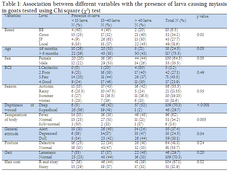
Table 1: Association between different variables with the presence of larva causing myiasis in goats tested using Chi square (χ2) test
Myiasis According to Risk Factors
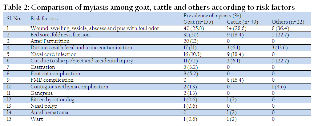
Wounds with foul odor, swelling, vesicle and abscess by bacterial contamination were the most crucial risk factors for myiasis. However, breach after delivery in cows, umbilical infection in new born kids, insanitary and germ–infested infrastructure of housing leads to bed sore were the most significant risk factors. Additionally, dirtiness and wetted surroundings (fleece, hair) with fecal and urine contamination, lack of aseptic surgery like castration with unsterile instruments (scissors, scalpel, forceps etc.), accidental injury (nail, barbed wire, glass etc.) as other major risk factors was evaluated. Furthermore, various diseases such as FMD (Foot and Mouth Disease), foot rot, contagious ecthyma, aural hematoma were complicated with myiasis. It was also experiential that, animals, were bitten by rat or dog (carnivores) create space on that biting place predisposed to myiasis. Amazingly, gangrene, wart, polyps were further recorded risk factors of myiasis (Table 2)
Comparison of Myiatic wound between Cattle and Goat
Among all wounds (Incised, septic, lacerated, punctured etc.), septic wounds were found more prominent than others (Cattle 69.4% and Goat 55.5%) while incised wound was recorded as persistent in goat than cattle (Cattle 6.1% and Goat 19.36%).
Myiasis According to Affected Body Regions
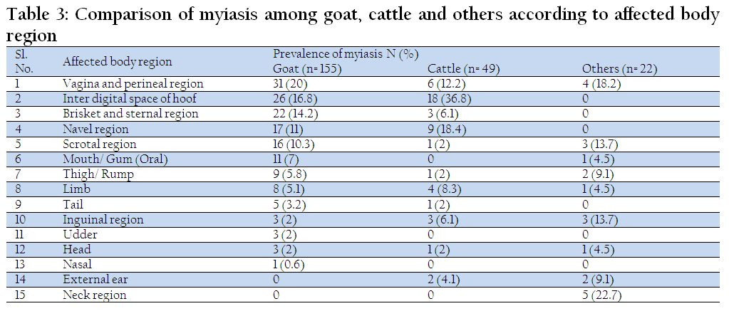
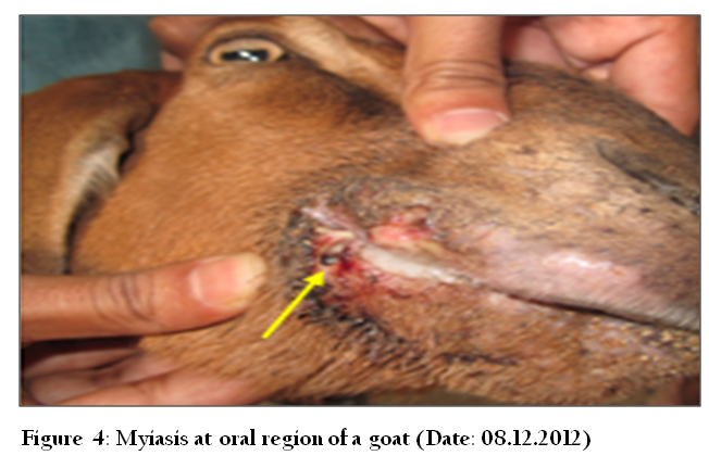
Myiasis was observed regardless of body area though vagina and perineal region, tail, inter digital space, brisket region, navel, scrotal region, inguinal region, udder, thigh/rump, limb, head region, mouth/gum, nasal, ear and neck region were recorded more vulnerable. Details of myiasis affected regions are illustrated in Table 3.
DISCUSSION
Overall prevalence of our study was 5.21% among 4338 cases which is comparable to Giangaspero et al. (2011), Alahmed (2004) who reported 3% out of 3129 in Italy, 2% out of 3712 in Riyadh Region respectively. However, Radfar and Hajmohammadi (2012), Shoorijeh et al. (2011), Gebremedhin EZ (2011), Arslan et al. (2009), Kara et al. (2005), Abo–Shehada et al. (2003) and Dorchies et al. (2000) found higher prevalence rate of myiasis. In the study, 226 cases were myiasis where goat 69%, cattle 22%, dog 7% and sheep 2% which is compared to Sergio et al. (2007) stating cattle and goat 46.4%, followed by dogs 15.3%, humans 14.7%, pigs 6%, horses 4% and sheep 1%. Apparent deviation is reflected the differences in the level of management, housing, cleanliness and as well as genetic variation in disease resistance breeds. Cross breed goat were commonly infested with myiasis (34.2%) than local (31.6%), Jamnapari (27.7%) and Black Bengal (6.5%) due to genetic variation and environment of tropics which is agreed by Kara et al. (2005) and Farkas et al. (1997); however, Cramer et al. (2002) found adult, light and short haired pure male dog breeds were mostly infested. In our study, females were significantly (p ≤ 0.05) infested with myiasis (64.5%) than male (35.5%) which had a similar finding with Radfar and Hajmohammadi (2012), but their relationship was insignificant. However, Orfanou et al. (2011), Abd El–Rahman (2010), Kara et al. (2005), Farkas et al. (1997) reported more cases in male. Shoorijeh et al. (2011) and Abo–Shehada et al. (2003) found same type of infection rate in both sexes. Poor to fatty animals were infested with myiasis, which is agreement with Gebremedhin (2011). Above 6 months old goats were more prone to myiasis (75.5%) (p ≤ 0.05) which is close to Rahman et al. (2009) where myiasis mostly occurs in cattle of over 2 years. However, different agreement by Kara et al. (2005) implicit infestation which is decreased with the age of cattle and Abo–Shehada et al. (2003) stating all age groups were equally infested. Paredes–Esquivel et al. (2012) found prevalence in lambs younger than 4 months was significantly affected (p ≤ 0.05) which was insignificant in adult sheep (p=0.081).
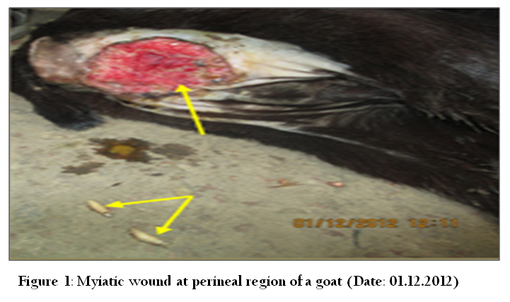

Figure 2: Myiasis at the scrotal region of a goat, occurred after 4 days of castration (Date: 21.11.2012)
Wounds, swelling, vesicle, abscess and pus with foul odor have been identified mostly as major predisposing factors. Septic wounds of different parts of body were more prominent than others (Goat 55.5% and Cattle 69.4%). Particularly deep wound significantly harboring huge maggots compared to superficial wounds (p ≤ 0.05) which is an agreement with Farkas and Hall (1998). Among other body parts, myiasis frequently occurs at vagina and perineal region (Goat 20%, cattle 12.2%); inter digital space (Goat 16.8%, Cattle 36.8%) which coincide with Rahman et al. (2009). Moreover, Giangaspero et al. (2011) found myiatic wound on vulva and prepuce of sheep; Gaglio et al. (2011) reported three cases of genital myiasis of a goat, ram and dog in Italy; Duro et al. (2007) stated umbilical myiasis in animal; Farkas et al. (1997) found wound myiasis on external genital organs (87%) of sheep in Hungary. Our study exposed other common sites as brisket (goat 14.2% and cattle 6.1%), navel (goat 11% and cattle 18.4%), scrotal (goat 10.3% and cattle 2%), tail, inguinal, udder, thigh/rump, limb, gum, nasal, horn, ear and neck which is similar findings with Cramer et al. (2002) in dog of southern zone of Rio de Janeiro municipality, Farkas et al. (2009) in dog of Al Hoceima, northern Morocco, Beth Knapp–Tyner, (2010) in deer fawn.

On the observation, dirtiness with fecal and urine contamination, poor housing infrastructure, floor with rough surface (brick, concrete) leads to bed sore and wound which is agreement with Bhola et al. (2012). Phillips (2009) confirmed sheep were predisposed to fly strike (cutaneous myiasis) where fleece was contaminated with feces or urine. Accidental injury and traumatic wound (nail, barbed wire, glass etc.) is another important risk factor causing myiasis. Trombetta et al. (2009) reported traumatic myiasis habitually in cattle, dogs and cats whereas Farkas and Hall (1998) found traumatic myiasis infestation >10% of animals at sheep, cattle and horses in Hungary. Additionally, Dik et al. (2012) found 22 traumatic myiasis in animals in Turkey; Ipek DN and Ipek P (2012) observed a facultative traumatic myiasis in domestic rabbit; Giangaspero et al. (2011) examined 10 traumatic myiasis in Italy. Scholtz et al. (2011) reported as presence or absence of dermatophilosis was the main predisposing factor for blowfly strike in sheep. Amazingly gangrene, wart, polyp are fascinating risk factors of myiasis.
Our study revealed that myiasis was found highest in autumn (55.5%) than cold (11.6%) due to prevailing warm and humid climate of tropic where Radfar and Hajmohammadi (2012) reported prevalence varied from 6.8% to 41.8% in August, 2007 to February, 2008 in South–eastern part of Iran; Paredes–Esquivel et al. (2012) recorded significant differences in prevalence in winter and autumn where fly activity held between May to June in the island of Majorca (Spain). In addition, Shoorijeh et al. (2011) found prevalence ranged from 6.6% in spring to 17.9% in winter in South Iran where Abd El–Rahman (2010) analyzed infestation rate in camel was significantly greater in colder (68.8%) than warmer (31%) in Western Libya. Similarly, Orfanou et al. (2011) found six cases from May to July and three cases from August to October in 163 dogs. Alem et al. (2010) described prevalence ranged from 77.7% to 98.8% from November to March in sheep and goats in Central Oromia; Arslan et al. (2009) reported prevalence of nasal myiasis was 54.3% in spring, 41% in summer, 28% in rainfall, 38.9% in winter and statistically significant differences among seasons (p ≤ 0.05) at north–eastern part of Turkey; Alahmed (2004) stated highest percentages of myiasis during Mar–May (60%) and Sept–Nov (31.5%) where temperature and relative humidity are optimum and infestation incidences were low (5% and 1.5% respectively) at dry hot season (Jun–August) and cold season (Dec–Feb) in Riyadh Region; Dorchies et al. (2000) found prevalence from 14.3% to 65% in Feb–Oct in sheep and 6.25% to 47.1% in Sept–Apr in goat in France; Farkas and Hall (1998) described myiasis season lasted from March to November where most cases were available in July and August at sheep, cattle and horses in Hungary; Amin et al. (1997) revealed high infestation rate in summer, followed by spring then autumn. However, Cramer et al. (2002) said no month of the year presented higher occurrence of myiasis cases.
CONCLUSION
Myiasis is the most familiar and widely distributed disease in Chittagong, Bangladesh. This research has addressed the problem of myiasis in Bangladesh, particularly in Chittagong, so that they will take necessary measures to make the problem subside, animal owners or rearers in avoiding and clinically managing them as well. Further widespread studies are suggested as molecular identification of species of flies and economic analysis caused by myiasis.
ACKNOWLEDGEMENT
The authors are highly grateful to the owners and veterinary clinicians, especially DR. Tarana Ahmed, Veterinary Surgeon, Patiya for her cordial cooperation participated in this study.
REFERENCES
Abd El–Rahman SS (2010). Prevalence and Pathology of Nasal Myiasis in Camels Slaughtered in El–Zawia Province–Western Libya: with a Reference to Thyroid Alteration and Renal Lipidosis. Global Veterinaria. 4 (2): 190–197.
Abo–Shehada MN, Batainah T, Abuharfeil N and Torgerson PR (2003). Oestrus ovis larval myiasis among goats in northern Jordan. Prev. Vet. Med. 59(1–2): 13–19.
http://dx.doi.org/10.1016/S0167-5877(03)00058-8
Alahmed AM (2004). Myiasis in sheep farms in Riyadh Region, Saudi Arabia. J. Egypt. Soc. Parasitol. 34(1): 153–160.
PMid:15125523
Alem F, Kumsa B and Degefu H (2010). Oestrus ovis larval myiasis among sheep and goats in Central Oromia, Ethiopia. Trop. Anim. Health Prod. 42(4): 697–703.
http://dx.doi.org/10.1007/s11250-009-9477-6
PMid:19882360
Amin AR, Shoukry A, Morsy TA and Mazyad SA (1997). Studies of wound myiasis among sheep and goats in North Sinai Governorate, Egypt. J. Egypt. Soc. Parasitol. 27(3): 719–737.
PMid:9425818
Arslan MO, Kara M and Gicik Y (2009). Epidemiology of Oestrus ovis infestations in sheep in Kars province of north–eastern Turkey. Trop. Anim. Health Prod. 41 (3): 299–305.
http://dx.doi.org/10.1007/s11250-008-9190-x
PMid:18523857
Beth Knapp–Tyner (2010). Deer Fawn Myiasis; Wildlife Rehabilitators of North Carolina. (http://www.ncwildliferehab.org/wordpress/programs/deer_fawn_myiasis.html) Accessed 25 January 2013
Bhola N, Jadhav A, Borle R, Adwani N, Khemka G and Jadhav P (2012). Primary Oral Myiasis: A Case Report in India. Case Reports in Dentistry Volume 2012, Article ID 734234, 4 pages.
Bolognia JL, Jorizzo JL and Rapini R (2008). Cutaneous myiasis. In: Dermatology. Vol 1, 2nd edn, Mosby Elsevier. 1300–1301pp.
Catts EP and Mullen GR (2002). Myiasis (Muscoidea, Oestroidea). Medical and Veterinary Entomology, Elseiver Sci. (USA). 317–348 pp.
http://dx.doi.org/10.1016/B978-012510451-7/50018-9
Cramer–Ribeiro BC, Sanavria A, Queiroz de Oliveira M, Silva de Souza F, Rocco F da S and Cardoso PG (2002). Inquiry of cases of myiasis by Dermatobia hominis in dogs of the southern zone of Rio de Janeiro municipality in 2000. Braz. J. Vet. Res. Anim. Sci. 39 (4): 176–180.
Dik B, Uslu U and Isik N (2012). Myiasis in Animals and Human beings in Turkey. Kafkas Univ. Vet. Fak. Derg. 18 (1): 37–42.
Dorchies P, Bergeaud JP, Tabouret G, Duranton C, Prevot F and Jacquiet P (2000). Prevalence and larval burden of Oestrus ovis (Linné 1761) in sheep and goats in northern Mediterranean region of France. Vet. Parasitol. 88(3–4): 269–273.
http://dx.doi.org/10.1016/S0304-4017(99)00215-0
Duro EA, Mariluis JC and Mulieri PR (2007). Umbilical myiasis in a human newborn. J. Perinatol. 27(4): 250–251.
http://dx.doi.org/10.1038/sj.jp.7211654
PMid:17377609
Farkas R and Hall MJ (1998). Prevalence of traumatic myiasis in Hungary: a questionnaire survey of veterinarians. Vet. Rec. 143(16): 440–443.
http://dx.doi.org/10.1136/vr.143.16.440
PMid:9823605
Farkas R, Hall MJR and Kelemen F (1997). Wound myiasis of sheep in Hungary. Veterinary Parasitology. 69 (1–2): 133–144.
http://dx.doi.org/10.1016/S0304-4017(96)01110-7
Farkas R, Hall MJR, Bouzagou AK, Lhor Y and Khallaayoune K (2009). Traumatic myiasis in dogs caused by Wohlfahrtia magnifica and its importance in the epidemiology of wohlfahrtiosis of livestock. Med. Vet. Entomol. 23(Suppl. 1): 80–85.
http://dx.doi.org/10.1111/j.1365-2915.2008.00772.x
PMid:19335833
Gaglio G, Brianti E, Abbene S and Giannetto S (2011). Genital myiasis by Wohlfahrtia magnifica (Diptera, Sarcophagidae) in Sicily (Italy). Parasitol. Res. 109(5): 1471–1474.
http://dx.doi.org/10.1007/s00436-011-2431-3
PMid:21541751
Gebremedhin EZ (2011). Prevalence of ovine and caprine oestrosis in Ambo, Ethiopia. Trop. Anim. Health Prod. 43(1): 265–270.
http://dx.doi.org/10.1007/s11250-010-9687-y
PMid:20725855
Gingaspero A, Traversa D, Trentini R, Scala A and Otranto D (2011). Traumatic myiasis by Wohlfahrtia magnifica in Italy. Vet. Parasitol. 175(1–2): 109–112.
http://dx.doi.org/10.1016/j.vetpar.2010.09.028
PMid:21030155
Ipek DNS and Ipek P (2012). A Case of Traumatic Myiasis in a Domestic Rabbit (Oryctolagus cuniculus) Caused By Lucilia sericata. Turkiye Parazitol. Derg. 36(1): 54–56.
http://dx.doi.org/10.5152/tpd.2012.14
PMid:22450925
Jelinek T, Nothdurft H, Rieder N and Loescher T (2000). Cutaneous Myiasis: review of 13cases of travelers returning from tropical countries. Int Dermatol. 39(9): 689–694.
John HK (1999). Screwworms: Be on the Lookout. In: Veterinary Medicine Extension. University of California, Davis Tulare CA 93274.
Kara M, Arslan M and Gicik Y (2005). The Prevalence of Bovine Hypodermosis in Kars Province, Turkey. Trop. Anim. Health Prod. 37 (8): 617–622.
http://dx.doi.org/10.1007/s11250-005-4291-2
PMid:16619878
Myiasis Wiki–vet (2011). Myiasis–causing flies, The Animal Health & Production Compendium (AHPC), published online by CABI during the OVAL Project. http://en.wikivet.net/Myiasis. Accessed 16 January 2013
Orfanou DC, Papadopoulos E, Cripps PJ, Athanasiou LV and Fthenakis GC (2011). Myiasis in a dog shelter in Greece: epidemiological and clinical features and therapeutic considerations. Vet. Parasitol. 181 (2–4): 374–378.
http://dx.doi.org/10.1016/j.vetpar.2011.04.006
PMid:21536388
Otranto D, Traversa D and Giangaspero A (2004). Myiasis caused by Oestridae: serological and molecular diagnosis. Parassitologia. 46 (1–2): 169–172.
PMid:15305710
Papadopoulos E, Himonas C and Boulard C (1997). The prevalence of bovine hypodermosis in Greece. Parassitologia. 39 (4): 431–433.
PMid:9802108
Paredes–Esquivel C, del Rio R, Monerris M, Borras D, Laglera LM and Miranda MA (2012). The influence of sheep age group on the seasonal prevalence of oestrosis in the island of Majorca (Spain). Vet. Parasitol. 186(3–4): 538–41.
http://dx.doi.org/10.1016/j.vetpar.2011.11.065
PMid:22186195
Phillips CJC (2009). A review of mulesing and other methods to control fly strike (cutaneous myiasis) in sheep. Universities federation for animal welfare, The Old School, Brewhouse Hill, Wheathampstead, Hertfordshire, UK, 18: 113–121. http://espace.library.uq.edu.au/eserv/UQ:233179/UQ233179_fulltext.pdf
Radfar MH and Hajmohammadi V (2012). Prevalence of goat warble fly, Przhevalskiana silenus in southeastern of Iran. Sci. Parasitol. 13 (2): 73–76.
Radostits OM, Gay CC, Blood DC and Hinchcliff KW (2000). Clinical examination and making a diagnosis. In: Veterinary Medicine, A textbook of the diseases of cattle, sheep, pigs, goats and horses. 9th edn. W B Saunders. 3–40 pp.
Rahman MA, Hossain MA and Alam MR (2009). Clinical evaluation of different treatment regimes for management of myiasis in cattle. Bangl. J. Vet. Med. 7 (2): 348 – 352.
Scholtz AJ, Cloete SW, du Toit E, van Wyk JB and van der Linde TCA (2011). Survey of the prevalence of blowfly strike and the control measures used in the Rûens area of the Western Cape Province of South Africa. J. S. Afr. Vet. Assoc. 82 (2): 107–15.
http://dx.doi.org/10.4102/jsava.v82i2.43
PMid:22135924
Sergio EB, José DE, Angel BC, Franklin C, Janina S, Sabina B and Enrique M (2007). Incidence of myiasis in Panama during the eradication of Cochliomyia hominivorax. Sección de Entomología Médica, Instituto Conmemorativo Gorgas de Estudios de la Salud, PO Box 0816–02593, Panamá.
Serra–Freire NM and Mello RP (2006). Entomologia & Acarologia na Medicina Veterinária. 1st edn. Editora L. F. Livros de Veterinária Ltd.
Sherman RA (2000). Wound myiasis in urban and suburban United States. Arch Intern Med 160: 204.
http://dx.doi.org/10.1001/archinte.160.13.2004
Shinohara EH, Martini MZ, Oliveira Neto HG and Takahashi A (2004). Oral myiasis treated with ivermectin: Case report. Braz Dent J. 15: 79–81.
http://dx.doi.org/10.1590/S0103-64402004000100015
PMid:15322651
Shoorijeh JS, Tamadon A, Negahban SH and Behjadi MA (2011). Prevalence of Oestrus ovis in goats of Shiraz, southern Iran. Vet. arhiv. 81 (1): 43–49.
Thrusfield MV (2005). Criteria for Success of Questionnaire. In: Veterinary Epidemiology. 3rd edn, Oxford, UK: Blackwell Science. 189–213pp.
Trombetta L, Oliva A, Galache V, Bava J and Troncoso A (2009). Cutaneous myiasis due to Cochliomyia hominivorax in a drug user. J. Infect. Dev. Ctries. 3(11): 873–876.
PMid:20061685


