Research Journal for Veterinary Practitioners
Case Report

Case Report of Rectourethral Fistula and Atresia Ani in a Day Old Rottweiler Puppy
Ziaullah1*, Hira Anjum1, Abid Hussain Shahzad2
1Pet Center, University of Veterinary and Animal Sciences, Lahore; 2College of Veterinary and Animal Sciences, Jhang-35200, Pakistan.
Abstract | Atresia ani with rectourethral fistula is a hereditary anomaly that influences the anal opening and rectum by development of an atypical association between the rectum and urethra. This anomaly was identified in a day old Rottweiler male Puppy. The presenting physical abnormalities included depression, dehydration, anal atresia and a discharge of watery feces from the vaginal opening. The puppy was in normal health conditions as owner has noticed the absence of anal opening. On clinical examination is was type II condition. Exploratory and corrective surgery does prove that besides type II atresia ani, it has rectourethral fistula which was corrected surgically.
Keywords | Rottweiler puppy, Atresia ani, Rectourethral fistula, Atypical case, Surgical correction
Received | March 31, 2020; Accepted | May 11, 2020; Published | June 04, 2020
*Correspondence | Ziaullah, Pet center, University of Veterinary and Animal Sciences, Lahore; Email: ziaullah.mughal@uvas.edu.pk
Citation | Ziaullah, Anjum H, Shahzad AH (2020). Case report of rectourethral fistula and atresia ani in a day old rottweiler puppy. Res J. Vet. Pract. 8(2): 20-22.
DOI | http://dx.doi.org/10.17582/journal.rjvp/2020/8.2.20.22
ISSN | 2308-2798
Copyright © 2020 Ziaullah et al. This is an open access article distributed under the Creative Commons Attribution License, which permits unrestricted use, distribution, and reproduction in any medium, provided the original work is properly cited.
Introduction
Hereditary anomalies related to anus and/or rectum origin in association with urogenital deformities are considered to be resulting from unusual embryonic development within the cloacal region. Cloacal opening is a common pathway for gastrointestinal, urinary, and reproductive tracts (Suess et al., 1982). Rectourtheral fistulae are linked with a malfunction of the urorectal fold to split up the developing cloaca properly at embryonic stages resulting in two separate openings for the urogenital and gastrointestinal tracts (Suess et al., 1982). Caudal part of the urogenital sinus develops into the urethra (Goulden et al., 1973). On the basis of literature reports it has been concluded that main reason for of fistula formation to be failure of the urorectal septum to completely divide the cloaca into an anterior urethrovesical segment and a posterior rectal segment. Similarly, atresia ani is also an inherited state that concerns the anal opening, rectum and may occur in both canines and felines. Numerous anatomical classes of atresia ani have been described ranging from type I – IV. Individuals with type I, otherwise termed imperforate anus, have a small covering over the anal opening, but the rectum ends as a blind pocket immediately cranial to the anal opening. Type II and I are alike, only the difference is the rectal pocket ends much more cranially to the anal opening in type II. In type III atresia ani, also called rectal atresia, rectum ends in a blind-ending pouch in the abdomen but distal rectum and anus are typical. Type IV atresia ani limited to females only and can occur with or without imperforate anus. It is distinguished by a persistent connection between the rectum and vagina (rectovaginal fistula) or urethra (rectourethral fistula; Ettinger and Feldman, 2017).
History and treatment
A day old Rottweiler male puppy was presented to Zia veterinary clinic (139-G, Commercial Area Defense Housing Authority Phase-I, Lahore) with the complaint of not passing stool from last 24 hours. The puppy was having normal vitals and good health condition. Body weight was 297g and body temperature 100.70F. On physical examination the anal opening was absent (Figure 1A). The urine passed by puppy was mixed with stool material. Emergency laprotomy was performed to correct the anomaly. The puppy was maintained under Isoflurane in general anesthesia. This general anesthesia was achieved by incremental method. Briefly 3 min of preoxygenation and then introduction of 0.5% vapor setting for 30-60 seconds and then 0.5% increment for the same period. After aseptic preparations, an incision was given on ventral midline through skin, subcutaneous tissue and linea alba into the body cavity. Urinary bladder and rectum were indentified visually. First the rectum was given an enterotomy incision and using a probe through it, external end of the rectum was identified in anal area and through stab incision the anal opening was made and skin flaps were sutured (Figure 1B).
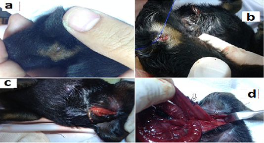
Figure 1: (A) a day old, male Rottweiler puppy with type II atresia ani (rectouretheral fistula). (B) Reconstruction through anoplasty in day old puppy with atresia ani type-II. (C) Closed abdominal incision showing that no sutures were placed on the skin. (D) Enterotomy incision closed, it was used to identify the anal opening and the contact point of rectum with the anus using a probe (arrow a) and Urethrorectal fistula being closed using absorbable suture material Vicryl 3/0 (arrow b).
The enterotomy incision was closed using absorbable sutures using Vicryl 4/0. The fistula was identified at the level of pubis. The pudendal artery, vein, and nerve were then identified and the fistula was located by using blunt dissection. The fistula was closed using absorbable suture material 3/0 Vicryl (Figure 1D). The abdominal incision was closed in two layers. The linea alba was closed with simple continuous sutures using absorbable suture material Vicryl 4/0, and buried simple continuous subcuticular sutures were placed with same suture material. No sutures were placed on the skin (Figure 1C). Pain medication and prophylactic antibiotic therapy was carried out for three days along with local wound dressing twice a day. Antibiotic therapy (Clavamox @10 mg/kg b.i.d.) continued for seven days. The puppy meanwhile was on mother feed alone and recovered uneventfully with functional anal opening.
Discussion
Rectouretheral fistula is believed to be developmental anomaly during embryonic life. It occurs due to the failure of the urorectal septum to split the cloacae into the urethrovesical and rectal sections. It may be classified from type I-IV depending upon the extent and involved organs. Type IV atresia ani is frequently linked with tenesmus and is diagnosed by the watery feces via the vagina or urethra with a characteristic perivulvar redness and infection. Diagnosis depends on the typical physical defects in affected individual with a concurrent history of tenesmus or the lack of ability to defecate normally. Only treatment of choice for atresia ani is surgical intervention. Prognosis is poor to guarded from attainment of normal functionality of rectum post treatment. Despite the type of abnormality, young and weak puppies often do not survive the procedure. Some affected individuals develop concurrent megacolon, which develop as a result of persistent fecal impaction which is irreversible condition, necessitating emergent management or subtotal colectomy (Ettinger and Feldman, 2017). Additionally, it is wise to examine the animal for further abnormalities when atresia ani is assumed. In literature, concurrent disorders, such as congenital hydrocephalus, have been documented (Suess et al., 1982). In one study, all of the three dogs with rectovaginal fistula and atresia ani had partial tail agenesis (Rahal et al., 2007).
According to previous literature this anomaly was more prevalent in male dogs but not reported in Rottweiler Puppy. Postoperative complication like tenesmus, fecal incontinence, and rectal prolapse have been described by many authors (Suess et al., 1982; Prassinos et al., 2003; Ellison and Papazoglou, 2011). Fecal incontinence, a common complication after surgery, may be transitory (Rahal et al., 2007), irregular or complete (Suess et al., 1982; Ellison and Papazoglou, 2011) and related to a congenital absence of functional external anal sphincter or surgical trauma to the sphincter muscle innervation during dissection (Aronson, 2003; Prassinos et al., 2003; Viana and Tobias, 2005; Ellison and Papazoglou, 2011). It may be secondary to surgical approach in dogs and takes longer time to resolve (Viana and Tobias, 2005; Rahal et al., 2007). Semitendinosus muscle flap technique was suggested as an option to improve anal function in a dog with atresia ani and rectovaginal fistula (Chambers and Rawlings, 1991). In current case, fortunately, recovery rate was high and no complications were faced except constipation after 3 days postoperative but issue was resolved in a week.
Authors Contribution
ZM and HA conducted the surgical procedure. ZM, HA and AHS drafted the manuscript. All authors edited the draft manuscript before final submission.
Conflict of interest
The authors have declared no conflict of interest.
References


