Research Journal for Veterinary Practitioners
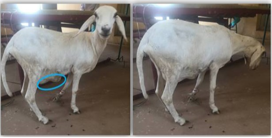
Patient on presentation showing the swelling on the ventral abdominal region.
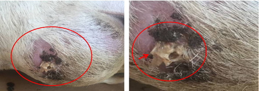
The patient on presentation showing the burst swelling on left lateral recombency. The circumscribed area shows the pus with protruding fragment of fetal bone (arrow head).
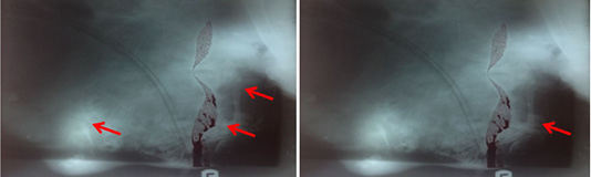
Lateral radiograph of the abdominal cavity showing the dismembered fetal bones. Red arrows showing dismembered fetal bones.
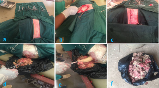
Showing the surgical procedure undertaken. (a) Triangular draping of the surgical field. (b) Incision into the abdomen via the paralumber fossa in order to exteriorize the punctured uterus for lavage and treatment. (c) Removal of the macerated fetus via the external opening. (e) Pulling of the skin remnant from the macerated fetus. (f) Complete macerated fetus removed from the uterus.


