Research Journal for Veterinary Practitioners
Case Report
Research Journal for Veterinary Practitioners 2 (1): 1 – 4A Case of Concurrent Inguinal Hernia and Vaginal Leiomyoma in a Bitch Successfully Treated Surgically
Kiranjeet Singh*, Deepti Bodh, Aswathy Gopinathan, Mohsina Abdul Muthalavi, Sangeetha Palakkara
*Corresponding author: ksuppli@yahoo.co.in
ARTICLE CITATION:
Singh K, Bodh D, Gopinathan A, Mohsina A and Sangeetha P (2014). A case of concurrent inguinal hernia and vaginal leiomyoma in a bitch successfully treated surgically. Res. j. vet. pract. 2 (1): 1 – 4
Received: 2013–10–23, Revised: 2013–11–18, Accepted: 2013–11–23
The electronic version of this article is the complete one and can be found online at
(http://www.nexusacademicpublishers.com/table_contents_detail/13/141/html)
which permits unrestricted use, distribution, and reproduction in any medium, provided the original work is properly cited
Abstract
An 11–year–old intact Pomerian bitch was presented to the Referral Veterinary Polyclinics, Indian Veterinary Research Institute, with a history of non–painful, non–reducible pendulous swelling (left side) on the caudal abdomen and a small perennial mass protruding through the vulva with no signs of discomfort. Abdominal radiography revealed protrusion of intestinal loops into the inguinal swelling confirming inguinal hernia Diagnosis of vaginal leiomyoma was confirmed by histopathology. Herniorrhaphy of the inguinal defect and surgical resection of the vaginal leiomyoma was performed under diazepam–ketamine anesthesia. No post–operative complications and reoccurrence of hernia and vaginal mass was reported up to one month after surgery.
INTRODUCTION
An inguinal hernia is a protrusion of an organ or part of an organ, fat or tissue through the inguinal ring, i.e. the region in the groin where the abdominal musculature meets the back legs. Factors potentially involved in the development of inguinal hernias may be anatomical, hormonal, and/or metabolic in nature; however the causes of inguinal herniation in small animals are poorly understood (Byers et al., 2007). Contents of inguinal hernia may include omentum, fat, ovary, uterus, small intestine, colon, bladder and spleen. These hernias are often chronic and do not cause clinical signs until pregnancy or pyometra develops (Fossum et al., 2002). Leiomyoma is a benign tumour originating from smooth muscle cells arising from any organ containing smooth muscle cells but the uterus and gastrointestinal tract are the most common sites (Yilmaz et al., 2009). Leiomyomas accounting for 85–90% of all canine uterine tumors, are the most common tumors of the tubular genitalia in dog and may be single or multiple, intraluminal or extraluminal and, in some unknown manner, endocrine dependent (Karagiannis et al., 2011). About 85% of leiomyomas occurring in the reproductive tract of a bitch arise from the vagina, vestibule and vulva (Bojrab MJ, 1975). The present case report describes the successful surgical management of concurrent inguinal hernia and vaginal leiomyoma in an elderly Pomerian bitch.
An 11 year old intact Pomerian bitch weighing 10 kg with a unilateral swelling in the inguinal region (prominent on left side) and a small mass protruding from vulva was presented to the Referral Veterinary Polyclinics of Indian Veterinary Research Institute. History revealed a long standing duration of swelling (approximately two years) in the inguinal region without any pain and discomfort to the bitch.
Clinical examination revealed normal heart rate, respiratory rate, rectal temperature and a general body condition. The bitch had whelped thrice with the last pup delivered two years back. A gradual increase in size of inguinal swelling had been observed since last estrus about 5–7 months back. A large, roughly orange sized, non–painful, non–reducible swelling involving abdominal mammary gland of left side was observed on physical examination near the inguinal region.
In the same bitch, another small mass was observed protruding through the vulva along with mild perineal swelling (Figure 1). The mass was present since 15–20 days but the bitch faced no difficulty in urination and defecation. Vaginal examination revealed an oval, smooth, firm mass arising from the region of ventral vaginal wall and extending into the vulvar opening. The skin and tissue overlying the mass was intact and appeared normal. However, the extent of attachment of mass to the vaginal wall could not be fully evaluated during examination due to the intact vaginal mucosa overlying the mass.

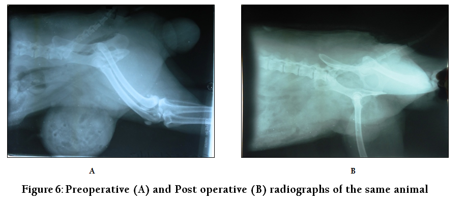
Lateral radiograph of caudal abdomen revealed two circumscribed swellings having soft tissue opacity in the inguinal and perenial region, respectively. The larger swelling in the inguinal region consisted of loops of intestine and gas pockets suggestive of inguinal herniation of intestine (Figure 6A). From the history, physical examination and diagnostic test results, inguinal herniation of intestine was confirmed and vagina leiomyoma was suspected.
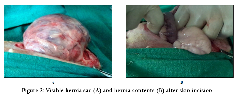
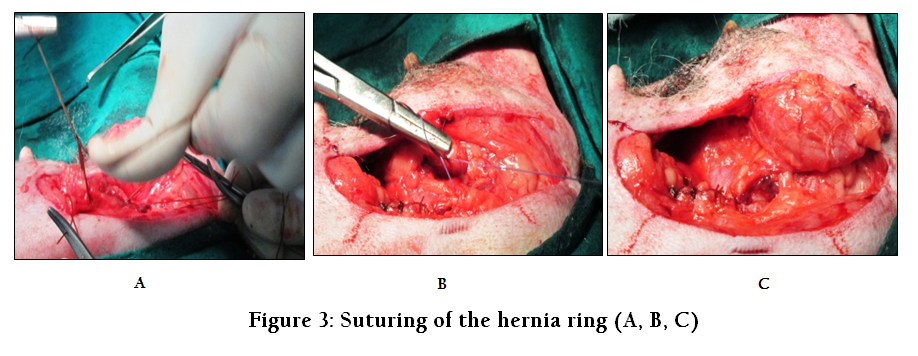
The bitch was premedicated by atropine (0.04 mg/kg IV) and five minutes later induced by diazepam (0.5 mg/kg IV) and ketamine (5 mg/kg IV), followed by endotracheal intubation. Anesthesia was maintained with diazepam–ketamine combination (1:1) administered intravenously. After preparation of the surgical site a skin incision was made directly over the swelling in the inguinal region just lateral to the groin fold. The subcutaneous tissue was undermined to allow exposure of the hernial ring and hernial sac, thereafter, an incision was made over the hernial sac and hernial contents comprising of omentum and small intestines were returned back into the abdominal cavity (Figure 2). The base of hernial sac was resected and the hernial ring was closed with No. 1–0 Vicryl in a simple interrupted suture pattern (Figure 3). The subcutaneous tissue was sutured in a simple continuous pattern using No. 0 Catgut to eliminate dead space and the skin incision was closed using No. 1–0 nylon in a cross mattress pattern.
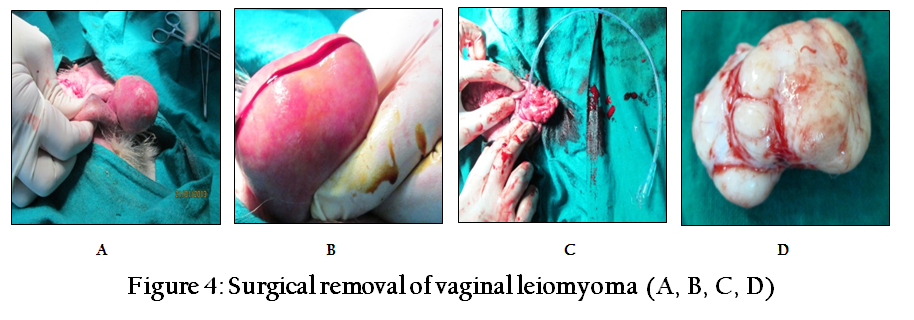
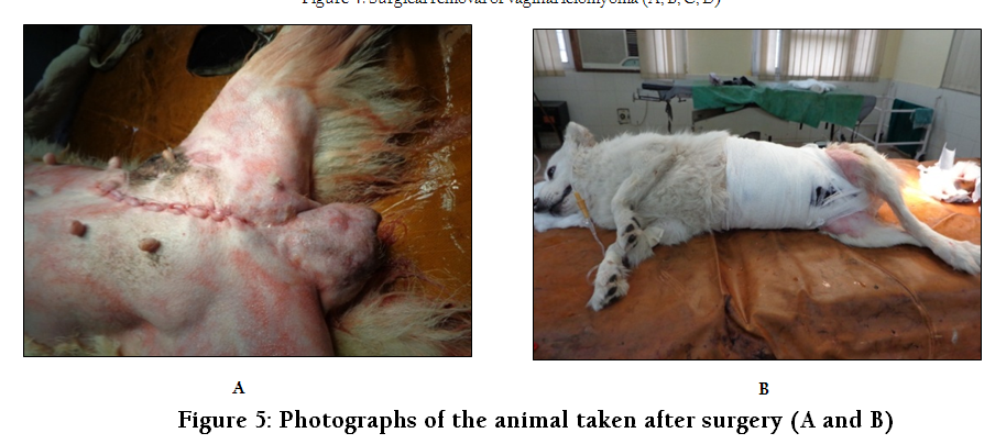
The mass in the perineal region was found to be smooth, oval, submucosal growth measuring approximately 6 cm in diameter (Figure 4). The mass was firm to touch and had a well defined stalk (3cm by 1 cm base) attached to the ventral vaginal wall. As the urethral orifice was very close to the pedicle of the mass, a size no.7 urinary catheter was placed in the urethra to help in defining and avoiding trauma to this structure (Figure 4C). The pedicle of the mass was then transected from base close to the urethra. After incision of the mass portions of it was sent for histopathological and cytological examination. The excision site was closed by approximating the mucosa of vaginal floor and submucosal dead space was occluded by simple interrupted suture pattern using No. 1–0 Chromic Catgut. Postoperative treatment consisted of Injection Ceftriaxone and Tablet Serratiopeptidase for five days, Injection Meloxicam and Tablet Rabeprazole for three days. Daily antiseptic dressing with Betadine (5% povidone iodine) and pressure bandaging on alternate days was advised. Sutures were removed on the 12th post operative day. The bitch made an uneventful recovery from anaesthesia with no postoperative complications.
Inguinal hernia occurs more often in middle aged intact bitches and may be caused by stretching of the abdominal muscles during pregnancy or atrophy of the abdominal wall due to old age (Waters et al, 1993). The clinical signs range from a painless inguinal mass to signs related to incarcerated or nonviable small intestine (Jahromi et al., 2009). As inguinal hernias typically appear during estrus or in pregnant bitches, estrogen is believed to play a major role in the development of these types of hernias (Fossum et al., 2002; Byers et al., 2007). Sex hormones may change the strength and character of the connective tissue, thereby weakening and enlarging the inguinal rings (Smeak et al., 2003). Unilateral inguinal hernias are more common than bilateral (Jahromi et al., 2009). Unilateral inguinal hernia was reported in this case without any involvement of contra lateral inguinal ring. Inguinal hernia can be diagnosed using radiography and ultrasonography (Abdin and Ramadan, 2001) as in the present case plain radiography revealed radiolucent intestinal loops protruding into the hernial sac. For the management of inguinal hernia in dogs three surgical approaches have been described and they include: midline approach (Smeak, 1993a), incision over the inguinal ring (Waters et al., 1993) and incision on the lateral aspect of hernia parallel to flank fold (Smeak, 1993a).
The surgical approach in this case involved an incision made on the lateral aspect of hernia parallel to the flank fold. As herniorrhaphy with simple interrupted or mattress suture has been found to be effective (Jahromi et al., 2009), same procedure was followed in this case. The hernial contents included omentum and intestine in the present case. Omentum is reported to be the most common organ present in canine inguinal hernias (Waters et al., 1993). Since inguinal hernia is mostly inherited, dogs that have had hernia or a surgical repair of hernia should not be used for breeding.
The most common types of tumors found in the genital tract of bitches include benign, smooth muscle tumors of the vagina and vulva. These tumors are variably referred to as leiomyomas, fibroleiomyomas, fibromas and polyps depending on the amount of connective tissue present (MacLachlan and Kennedy, 2002). The diagnosis of vaginal leiomyomas can be done histopathologically. In animals aetiology of leiomyomas is not fully understood but some studies suggest that hormones have an influence on their tumorigenesis (Miller et al., 2003). Vaginal leiomyomas are best treated surgically (Klein 2001) but the condition can recur due to hormonal influence (Miettinen and Fetsch, 2006). Therefore, ovariohysterectomy is advised at an early age to prevent occurrence of leiomyomas in female dogs.
Complications can occur after surgical repair of inguinal hernias as well as after resection of vaginal leiomyomas. Incisional infection, wound dehiscence, seroma, excessive postoperative swelling and peritonitis are some of the complications seen after surgical treatment for inguinal hernia in dogs (Jahromi et al., 2009). Prognosis was evaluated to be good in this case due to the absence of incarceration and intestinal perforation or leakage. Iatrogenic damage to the urethra or accidental injury to other perineal structures can occur during resection of vaginal mass while urine retention, incontinence and urethral obstruction may be noticed after resection. However, bitch in the present case did not show any of the postoperative complications or reoccurrence of hernia or vaginal leiomyoma till one month after the surgery.
REFERENCES
Abdin–Bey MR and Ramadan RO (2001). Retrospective Study of Hernias in Goats. Scientific Journal of King Faisal University (Basic and Applied Sciences). 2 (1):77–81.
Bojrab MJ (1975). Current techniques in small animal surgery I. Lea & Febiger. Philadelphia, 248 – 254.
Byers CG, Williams JE, Saylor DK (2007). Pyometra with inguinal herniation of the left uterine horn and omentum in a Beagle dog. J.Vet. Emerg. Crit. Care. 17: 86–92.
http://dx.doi.org/10.1111/j.1476-4431.2006.00212.x
Fossum TW, Choyl SH, Doland AH, Ann LJ, Howard BS, Michae DW (2002). Small Animal Surgery. 2nd ed. Mosby Inc Missouri, 261–267.
Jahromi AR, Nazhwani SD, Gandmani MJ, and Mehrshad S (2009). Concurrent bilateral inguinal and umbilical hernias in a bitch: a case report, Vet. Arhiv. 79 (5): 517–522.
Karagiannis GS, Pelekanis M, Loukopoulos P, Haris N, Ververidis HN and Kaldrymidou E (2011). Canine Uterine Leiomyoma with Epithelial Tissue Foci, Adenomyosis, and Cystic Endometrial Hyperplasia. Case Reports in Veterinary Medicine. Volume 2011 (2011), Article ID 901874, 4 pages. http://dx.doi.org/10.1155/2011/901874
http://dx.doi.org/10.1155/2011/901874
Klein MK (2001). Tumors of the female reproductive system. In: S.J. Withrow and E.G. MacEwen (eds), Small Animal Clinical Oncology. W. B. Saunders, Philadelphia, PA, USA, 445–454.
MacLachlan NJ, Kennedy PC (2002). Tumors of the genital system. In: D.J. Meuten (ed.), Tumors in domestic animals, 4th ed., Iowa State University Press, Ames, IA, 547–573.
http://dx.doi.org/10.1002/9780470376928.ch11
Miettinen M, Fetsch JF (2006). Evaluation of biological potential of smooth muscle tumors: Review. Histopathology. 48(1): 97.
http://dx.doi.org/10.1111/j.1365-2559.2005.02292.x
PMid:16359541
Miller MA, RamosVara JA, Dickerson MF, Johnson GC, Pace LW, Kreeger JM, Turnquist SE and Turk JR (2003). Uterine neoplasia in 13 cats. J. Vet. Diagn. Invest. 15: 575522
http://dx.doi.org/10.1177/104063870301500602
Smeak DD (1993a). Abdominal hernias. In: Slatter D. (ed.), Textbook of Small Animal Surgery, 2nd ed., WB Saunders, Philadelphia U.S.A. volume 1: 433–454.
Smeak DD (2003). Abdominal Hernias. In: Slatter D. (ed.), Text Book of Small Animal Surgery. 3rd ed., W B. Sunders, Philadelphia, U.S.A, 452–455.
Waters DJ, Roy RG, Stone EA (1993). Retrospective study of inguinal hernia in 35 dogs. Vet. Surg. 22, 44–49.
http://dx.doi.org/10.1111/j.1532-950X.1993.tb00367.x
Yilmaz R, Akkoc A, Ozyigit MO (2009). Ileal leiomyoma in a captive zebra (Equus burchelli). Turk. J. Vet. Anim. Sci. 33(5): 443–446


