Research Journal for Veterinary Practitioners
Research Article
Res. J. Vet. Pract. 7(3): 67-73
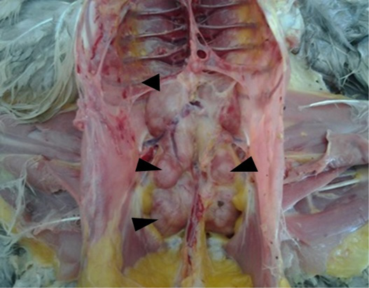
Figure 1
Renal tumor nodules (arrows) in a free-range pullet suspected of Marek’s disease
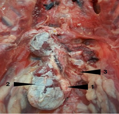
Figure 2
Visceral gout and urolithiasis (1) in a layer. Note hypertrophy (2) of the right kidney and the spectacular atrophy of the left kidney (3)
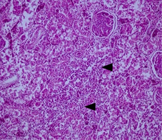
Figure 3
Kidney: acute interstitial nephritis in a layer suspected of IB. Note the marked infiltration by mononuclear cells in the interstitial space of renal tubules (arrows) (HEX200).
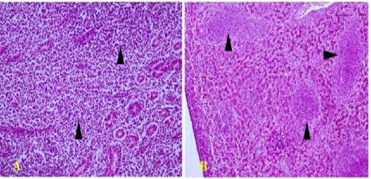
Figure 4
Polymorphic lympho-plasmocytic tumor infiltration in the kidney (arrows) (A; HEX200) and the liver (arrows) (B; HEX100)
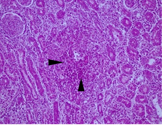
Figure 5
Kidney: necrotizing nephritis in a broiler suspected of salmonellosis (arrow) (HEX200)


