Research Journal for Veterinary Practitioners
Case Report

Arthrogryposis Multiplex Congenita and Perosomus Acaudatus Complicated with Atresia Ani in a Buffalo Calf
Khan Sharun*, Mohammed Arif Basha, Mudasir Ahmad Shah, Naveen Kumar Verma, Amarpal
Division of Surgery, ICAR-Indian Veterinary Research Institute, Izatnagar, Bareilly, Uttar Pradesh - 243122, India.
Abstract | The purpose of the present case report is to put on records an unusual and rare case of multiple congenital anomalies involving the central nervous system, musculoskeletal and gastrointestinal system in a one day old male Murrah buffalo calf. The buffalo calf was presented to Referral Veterinary Polyclinic, Indian Veterinary Research Institute, Izatnagar with the history of inability to stand and passage of small quantity of feces since birth through a punctured opening made by owner at perineal region. Physical examination of the calf revealed the absence of tail along with flexed and malformed limbs and incomplete anal opening. Radiographic examination of spine revealed the absence of sacral and coccygeal vertebrae along with malformed thoracolumbar vertebrae. On the basis ofhistory and clinical examination, the case was diagnosed asarthrogryposis multiplex congenita and perosomus acaudatus complicated with atresia ani type I (imperforate anus). The calf was operated for the correction of atresia ani. Postoperatively the calf was treated with nervine tonics to regain its bowel activity. However the calf died after a month following operation.
Keywords | Buffalo calf, Arthrogryposis multiplex congenita, Perosomus acaudatus, Atresia ani
Editor | Muhammad Abubakar, National Veterinary Laboratories, Islamabad, Pakistan.
Received | January 28, 2019; Accepted | February 17, 2019; Published | March 15, 2019
*Correspondence | Khan Sharun, Division of Surgery, ICAR-Indian Veterinary Research Institute, Izatnagar, Bareilly, Uttar Pradesh - 243122, India; Email: sharunkhansk@gmail.com
Citation | Sharun K, Basha MA, Shah MA, Verma NK, Amarpal (2019). Arthrogryposis multiplex congenita and perosomus acaudatus complicated with atresia ani in a buffalo calf. Res. J. Vet. Pract. 7(1): 35-38.
DOI | http://dx.doi.org/10.17582/journal.rjvp/2019/7.1.35.38
ISSN (Online) | 2308-2798
Copyright © 2019 Sharun et al. This is an open access article distributed under the Creative Commons Attribution License, which permits unrestricted use, distribution, and reproduction in any medium, provided the original work is properly cited.
Introduction
Congenital malformations occur when the genetic variants pass from parents to progeny. This may result in great economic losses to the breeders either due to death of the animal or damage to its reproductive and productive ability. Arthrogryposis is defined as a congenital disease characterized by non-progressive joint contractures that affect fore or hind limbs or the vertebral column, leading to varying degrees of flexion or limiting the extension (Albarella et al., 2017). The extent of malformations may vary depending upon the involvement of one, two, three, or all four limbs and the axial skeleton. Usually in mild cases there will be involvement of two legs, mostly the hind legs which will be in the flexed position (Jubb et al., 1993). Marcolongo-Pereira et al. (2010) reported the occurrence of congenital defects in Murrah buffalo as 7.54%. Among them, the most common defects were arthrogryposis, myotonia, and mechano-bullous genodermatoses.
Perosomus acaudatus is a congenital defect with the complete absence of the tailand is most common in Holstein cattle (Radostits et al., 2000). This condition is of either hereditary or congenital origin and mostly occurs due to the crossing between different breeds of cattle (Bähr and Distl, 2005).
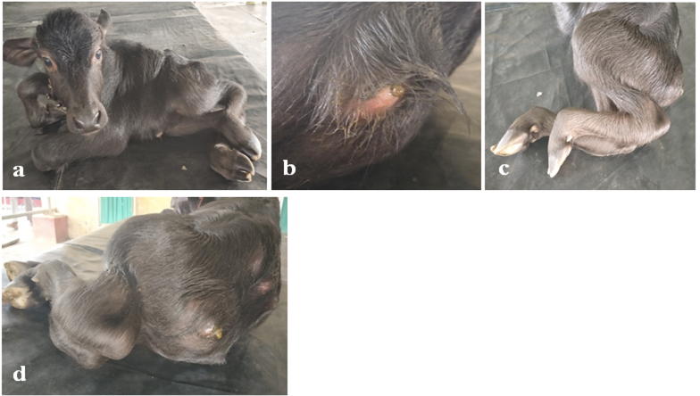
Figure 1: (a) Buffalo calf with flexed and malformed limbs, (b) Incomplete anal opening, (c) Flexed hind limbs involving multiple joints with muscle wasting indicating arthrogryposis multiplex congenita, (d) The complete absence of tail indicating perosomus acaudatus
The present study describes a rare case of arthrogryposis multiplex congenita and perosomus acaudatus complicated with atresia ani type I (imperforate anus) in a buffalo calf.
Case History and Clinical Examination
A one-day old male Murrah buffalo calf was presented to Referral Veterinary Polyclinic, Indian Veterinary Research Institute, Izatnagar with history of inability to stand and partial passage of feces since birth. On clinical examination, the calf had the absence of tail along with malformed fore and hind limbs and incomplete anal opening but was still active and alert. The animal was found unable to get up because of flexed and malformed limbs. On abdominal compression, a bulge was observed at the anal region followed by passage of fecal material through a small opening made by the owner using a pocket knife. Radiographic examination confirmed the absence of sacral and coccygeal vertebrae along with malformed thoracolumbar vertebrae (Figure 2). Enlarged rectum filled with gas and fecal material was also identified in the radiograph in dorsal pelvic cavity. On the basis of history and clinical examination, the case was diagnosed as arthrogryposis multiplex congenita and perosomus acaudatus complicated with atresia ani type I (Figure 1). It was decided to attempt an emergency surgery for the correction of type I atresia ani.
Surgical Technique
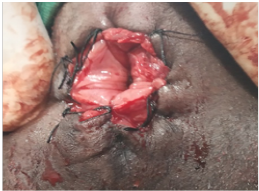
After aseptic site preparation, local anesthesia was achieved by infiltration of 2% lignocaine hydrochloride at the proposed incision site. Thereafter, the opening made by the owner was extended in a cruciate pattern without damaging the perineal muscles. The skin edges were trimmed to make it circular. The anal opening was reconstructed by suturing the rectal mucosa to the external skin by braided silk suture No. 1 using simple interrupted suture pattern (Figure 3).
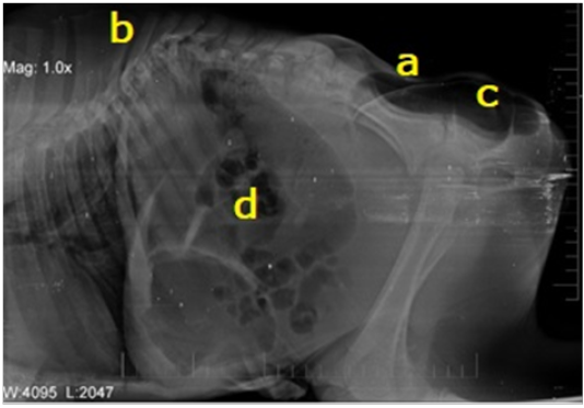
Figure 2: Right lateral abdominal radiograph showing (a) Complete absence of sacral and coccygeal vertebrae. (b) Malformation of thoracic and lumbar vertebrae. (c) Enlarged, gas and fecal matter filled rectum at the region of coccygeal vertebrae. (d) Gas-filled dilated intestinal loops
Post-operative Care
Postoperatively, injection Ceftriaxone (10 mg/ kg body weight, I/M) for five days, injection meloxicam (0.3 mg/kg body weight, I/M) for three days were administered. Injection Methylcobalamin (2500 mcg, I/M) for 2 weeks was administered to improve the bowel activity. The calf survived for one month and medical management helped to improve the quality of life of the calf.
Result
It was found that even after reconstruction of the anal opening, the animal did not pass faeces indicating functional obstruction of the distal gastrointestinal tract. The animal succumbed to death one month after the surgery.
Discussion
Congenital malformations are mainly due to a genetic cause. The development of an anomaly involves a complex mechanism hence, the causative agent resulting in congenital anomaly remains unknown (Kacar et al., 2008). Curly calf syndrome (Arthrogryposis Multiplex) is a disorder that emerged in Angus cattle in 2008 in Australia involving congenital contracture and fixation of several joints thus the term multiplex was given (Windsor et al., 2011). Arthrogryposis had varied etiology which includes physical limitation of in utero movementscausing hypokinesia or akinesia syndrome in the fetus and maternal illness. Intrauterine viral infections i.e. Akabane virus and Aino virus, toxins like Lupines sericus and Lupinus caudatus and genetic disorders of the fetus are also some of the causes (Agerholm et al., 2015). Leipold et al. (1970) reported the occurrence of arthrogryposis associated with palatoschisis in several breeds of cattle. Arthrogryposis is also associated with some other anomalies such asbrachygnathia, scoliosis, lordosis, kyphosis, hydranencephaly, and torticollis (Kacar et al., 2008). However the present case was further complicated by malformation of vertebral column, absence of sacral and coccygeal vertebrae and atresia ani.
Newborn animals which are affected with arthrogryposis are mostly stillborn or die within few days or weeks after birth. It is important to note that, though the animal had multiple congenital anomalies, it was active but lacked mobility which affected its ability to feed by itself. Lotfi and Shahryar (2009) reported a higher occurrence of Anury (absence of tail) in female dairy cattle than in male cattle. However, in the present case, the sex of the buffalo calf was male. From the radiographic examination, it was evident that the intestinal loops were gas filled and dilated, though animal passed scanty feces indicating some functional obstruction. It plausibly be due to the absence and/or damage to spinal nerves due to malformed vertebral column. Damage to the spinal cord due to malformed vertebrae along with the absence of terminal part of spinal cord may also affect the evacuation of bowel contents resulting in paralytic ileus. This might be the reason why the animal was not able to pass feces even when atresia ani was corrected surgically.
Identification of the cause for arthrogryposis and associated congenital anomalies are possible in a population but when it comes to sporadic cases of congenital anomalies such as the present case in buffalo calf, the etiology will remain unknown. Few authors have reported the occurrence of atresia ani associated with perosomus acaudatus in different species, calf (Reji et al., 2010), camel (Anwar et al., 2012), but very few literatures are present which reports the occurrence of such complicated congenital anomalies in buffalo. This paper reports the first ever case of atresia ani in a live buffalo calf having arthrogryposis multiplex congenita and perosomus acaudatus, which survived for one month after emergency surgery.
Conflict of interest
The authors declare that they have no conflict of interest.
Ethical approval
This article does not contain any studies with human or animal participants performed by any of the authors. All protocols followed were as per the guidelines from the standard textbooks in Veterinary Medicine and Surgery and were ethical.
References


