Research Journal for Veterinary Practitioners
Case Report
Res. J. Vet. Pract. 6(2): 14-19
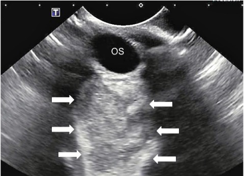
Figure 1
Ocular ultrasound of the left eye demonstrating a large hyperechoic retrobulbar space occupying lesion.
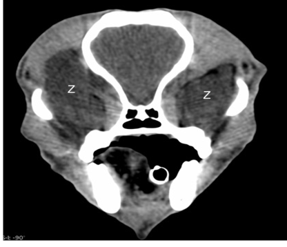
Figure 2
Pre-contrast computed tomographic image in a soft tissue window showing bilateral retrobulbar masses (Z). Both masses are hypodense and poorly defined and larger on the right (left side of image).
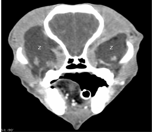
Figure 3
Post-contrast computed tomographic image in a soft tissue window showing the bilateral retrobulbar masses (Z) to have no enhancement.


