Research Journal for Veterinary Practitioners
Case Report

Differential Diagnosis of Orf from Peste Des Petits Ruminants; An Example from Field
Shumaila Manzoor1,2,*, Sami Ullah Khan Bahadur3, Usman Talib3, Muhammad Javed Arshad1, Muhammad Abubakar1, Aamer Bin Zahur1
1National Veterinary Laboratory, Park Road, Islamabad, Pakistan; 2FAO FMD and PPR project, NARC Premises, Park Road, Islamabad, Pakistan; 3University of Agriculture, Faisalabad, Pakistan.
Abstract | In a herd of 25 goats, a disease outbreak was reported in surrounding area of Islamabad. On the basis of clinical signs of oral lesions and very minute ocular and nasal discharge in 6 animals but diarrhea and difficult breathing was only in single animal, disease was suspected as Peste Des Petits Ruminants (PPR) by local veterinary practitioner. Animals were vaccinated for PPRV and swab samples were collected for further investigation. Samples were found negative for PPRV when tested through Penside and s-ELISA. Further physical examination revealed scabby lesions on face, papules at head and ear with normal respiration and heart rate. Hematological examination indicated lymphocytosis. On the basis of clinical signs, lymphocytosis and negative laboratory results for PPRV disease, outbreak was diagnosed as Orf. Animals were treated with parenteral inj. Tribressin®, inj. Vetafenac® and inj. Cyanocobe® along with topical applications at lesions. Animals recovered completely after 3 weeks of treatment intervention. It is suggestive to give more stress on differentially diagnosis of PPRV while working towards its progressive control leading to possible eradication.
Keywords | Contagious ecthyma, Orf, Peste Des Petits ruminats, Differential diagnosis
Editor | Muhammad Abubakar, National Veterinary Laboratories, Islamabad, Pakistan.
Received | April 04, 2018; Accepted | May 11, 2018; Published | August 30, 2018
*Correspondence | Shumaila Manzoor, National Veterinary Laboratory, Park Road, Islamabad, Pakistan; Email: shamaleo@hotmail.com
Citation | Manzoor S, Bahadur SUK, Talib U, Arshad MJ, Abubakar M, Zahur AB (2018). Differential diagnosis of orf from peste des petits ruminants; an example from field. Res. J. Vet. Pract. 6(2): 10-13.
DOI | http://dx.doi.org/10.17582/journal.rjvp/2018/6.2.10.13
ISSN (Online) | 2308-2798
Copyright © 2018 Manzoor et al. This is an open access article distributed under the Creative Commons Attribution License, which permits unrestricted use, distribution, and reproduction in any medium, provided the original work is properly cited.
Introduction
Orf is a viral disease of skin that is contagious in nature, having zoonotic potential. Primarily, it is a disease of small ruminants but wild and domesticated animals may also be affected. Disease is also named as contagious ecthyma, scabby mouth or contagious pustular dermatitis, which depicts that most often, target site is skin of mouth but other areas of skin may also be involved like udder, ear, head and foot (Kumar et al., 2015). Contagious ecthyma or sore mouth is caused by parapoxvirus (Housawi, 2002) which belongs to family poxviridae and subfamily Chordopoxvirinae (infecting vertebrates) (Friederichs et al., 2015). Pseudocowpox and Bovine popular stomatitis virus also belong to same genus of parapoxvirus.
Virus is transmitted through direct contact, sharing feeding and water troughs. Iatrogenic transmission is also possible (Hajkazemi et al., 2016). Contagious Ecthyma is a zoonotic problem and virus is transmitted from diseased animals to humans (Huerter et al., 1991). Disease is characterized by the presence of papules, vesicles or pustules and subsequently scabs on face skin. Orf has more destructive effects in goats than in sheep (Nandi et al., 2011). Differential diagnosis of disease is to be made with Foot and Mouth Disease, Bluetongue and Peste Des Petits Ruminants (PPR) (Kitching, 2001). Orf virus has primarily affinity for oral and perilabial area to cause erythematous and ulcerative papules (Kumar et al., 2015) but Peste Des Petits Ruminants (PPR) causes necrotic and erosive stomatitis (Abubakar et al., 2015). Such type of similarity in site of lesions is responsible for incorrect diagnosis of orf as PPR in the field. This misdiagnosis of Orf, having zoonotic potential leads to transmission of disease from affected animals to humans (Koufakis et al., 2014; Sanchez et al., 1985; Mohr and Katz, 1989).
Disease transmission to human is also possible by having any abrasion and cutting infected meat (Maor, 2017). Veterinarian, farmers and butchers are the people who are at risk if they do not handle animals with care, considering Orf as PPR. In this outbreak, disease was tentatively diagnosed as PPR by veterinary assistant but later on was confirmed as Orf. So, objective of this report is to differentiate between Orf and PPR and stress for differential diagnosis of PPR.
Case Presentation
This study was conducted in an area of Islamabad, where there was a suspected outbreak of PPR. A total of 5 Oral swab samples were received at NVL for the confirmation of PPR as diagnosed by a Veterinary Assistant. Anamnesis revealed that farmer introduced four new animals from lower Punjab in a very near past. On the basis of signs that were oral lesions and very minute ocular and nasal discharges and diarrhea, PPR was diagnosed tentatively and all animals were vaccinated for PPR by Veterinary Assistant but lab tests, Penside test and s-ELISA, were negative for PPRV. On the very next day, goat with signs of difficult breathing and diarrhea was died. This situation inclined to visit farm and 3 new incidences were found. Physical examination during visit showed scabby lesions at mouth area (Figure 1). One animal had papules on head and ear (Figure 2) Nictitating membranes were normal with no keratitis but very minute ocular discharge. Respiration rate and heart rate was also in normal range in all animals but submandibular lymph nodes were swollen. Anorexic condition with normal water drinking prevailed with no diarrhea in all diseased animals. As ketoprofen was being administered to all diseased animals prescribed by Veterinary Assistant, rectal temperature was in normal range.
Diagnosis
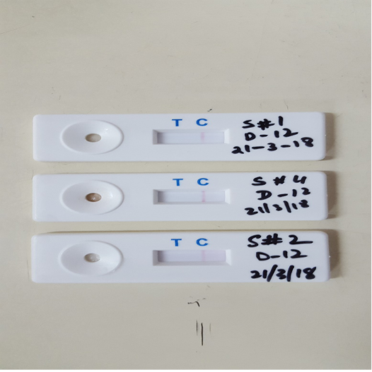
Oral swabs tested through Penside field test for PPRV infection (The Pirbright Institute Diagnostics ®) showed negative results for PPR (Figure 3). This is a simple dot ELISA for the detection of PPR antigen from tissue homogenate or swabs but having less sensitivity and specificity as compared to s-ELISA (Balamurugan, 2014). For confirmatory diagnosis, Sandwich ELISA for the detection of PPR virus (through ID Screen ® PPR Antigen Capture kit) was performed. This also showed negative results for PPR virus with S/P%=0.2 (<20%).
On the visit to farm, blood was collected from diseased animals for hematological examination. Blood smear stained with field stains examined under 100X revealed lymhocytosis. Hemoglobin level determined by Sahli’s apparatus, was found to be between ranges of 7.5-8.5 g/dL. Lymphocytosis inclined to consider disease as viral but was confirmed negative for Peste Des Petits Ruminants PPR as described. On the basis of clinical signs, diagnosis for PPR and hematological examination, disease outbreak was diagnosed as Orf or contagious ecthyma.
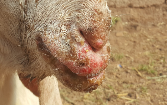
Figure 1: Scabby lesions at mouth area
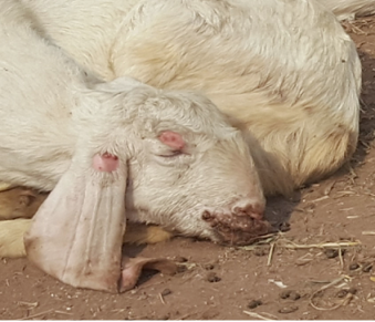
Figure 2: Papules on head and ear
Treatment
There is no specific treatment for contagious ecthyma (ORF) but supportive therapy is proved beneficial. Mortality is increased due to secondary bacterial infections (Abbas and Mughal, 2014). Diseased animals were prescribed with inj. Tribrissen® 3 ml for large animals and 2 ml for smaller OID through IM route for 4 days to combat any secondary infection, inj Meloxicam (Vetafenac®) for pyrexia with dose rate of 0.5 mg/kg and Inj. Cyanocobe® for better metabolism. Owner was advised (i) to give ORS dissolved water to resuscitate dehydration and (ii) to keep healthy animals separate from diseased. Somogel® and Bonjela® after mixing with Boric acid were prescribed to use topically on oral lesions so animals could continue to feed because anorexia due to pain of lesion is cause of death in Orf. This treatment worked and animals recovered after 3 weeks completely.
Discussion
Peste Des Petits Ruminants (PPR) and Orf are viral diseases, caused by PPR virus and parapoxvirus, respectively. PPR virus belongs to genus morbillivirus and family paramyxoviridae. PPR is characterized by pyrexia, ocular and nasal discharge that is serous but changes to purulent form, Diarrhea and Broncho-pneumonia. Oral lesions include necrosis and erosion of gums and buccal mucosa but Orf has erythematous and ulcerative papules in perilabial area. These papules and pustules are changed to scabs. Pneumonia, diarrhea, severe nasal and ocular discharge, were not seen in this outbreak except in one goat. In severe cases of Orf, pneumonia is followed by erosive stomatitis and gastroenteritis (Kumar et al., 2015) so animal died having signs of oral lesions, difficult breathing and diarrhea could have more severe disease.
Mortality rate is another feature that differentiates PPR from Orf. Mortality rate in case of PPR reaches up-to 100 % (Abubakar et al., 2008) while in Orf it is usually less than 1% but 20 % in only case of secondary bacterial infections (Abbas and Mughal, 2014). Similarly, case fatality of PPR in both sheep and goat is found to be >40 % and that of Orf is <1%. Abortion (28-45 %) at any stage of gestational period, is also associated with PPR outbreaks (Abubakar et al., 2008) while no such report found in case of Orf. Orf can be complicated by bacterial infections like Dermatophilus congolensis in foot lesions, maggots or screw-worms.
As Orf was diagnosed as PPR in this outbreak meanwhile it is also stated that PPR is sometime misdiagnosed as contagious ecthyma (Abubakar, 2016). This shows ambiguity related clinically diagnosing PPR and Orf. For confirmatory diagnosis of PPR, conventional tests like Penside, HA/HI, c-ELISA, Immuno-capture ELISA and molecular test RT PCR are choice (Abubakar, 2011). Orf can be diagnosed through examining scabs under electron microscope, histopathology, CFT and ELISA but under electron-microscope parapoxvirus of Orf can’t be differentiated from other parapoxviruses.
Animals suspected for Orf should be handled after wearing proper dress and gloves. Direct contact with affected animals is mode of transmission of disease from animals to humans. In human, Orf causes formation of pustules and papules mostly in arm region. In humans, disease is self-limiting but its immunosuppressive effect could make prone humans to secondary bacterial infections. There is need to make professionals and farmers about zoonotic disease of Orf to differentially diagnose Orf and PPR.
Acknowledgments
Authors want to acknowledge the support of FAO FMD and PPR project and the staff working in virology section, National Veterinary Laboratory.
Conflict of Interest
No conflict of interest is declared by Author(s).
Authors Contribution
SM and MA conceived the idea and drafted the skeleton of paper. SUKB and UT collected the samples. SM, SUKB and UT were involved in sample testing. SUKB and UT drafted the details of the manuscript. SM and MA did the final checking. ABZ and MJA did proof reading and all authors read and approved the final manuscript.
References


