Journal of Animal Health and Production
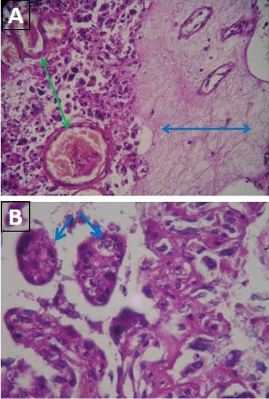
Histopathological sections in placentome of infected dam: A) Fibrinous precipitation (green arrow), polymorphonuclear cells infiltration and blood vessels congestion (blue arrow) (H&E stain x 40); B) Presence of pseudocyst containing several bradyzoites (blue arrow) (H&E stain x40)
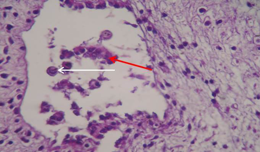
Showing section of brain of aborted fetus. Large necrotic cavities containing tissue debris indicating areas of encephalomalacia (Red arrow) with number of degenerating astrocysts (White arrow) (H&E stain x40)
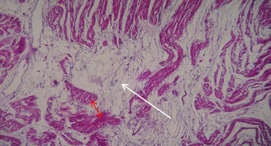
Showing section of heart of aborted fetus. Massive fragmentation and separation of myocardial bundles with intra muscular edema (White arrow) resulting in severe atrophy of survival muscle bundles (Red arrow) (H&E stain x40)
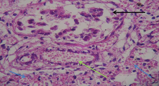
Showing section of lung of aborted fetus. Severe bloods vessels congestion (green arrow) associated with interstitial hemorrhage (blue arrow) with presence of pseudocysts containing several bradyzoites (black arrow) (H&E stain x40)


