Journal of Animal Health and Production
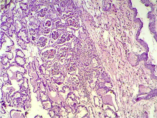
Mammary gland section shows sever destruction of mammary gland with abundant of inflammatory cells infiltration (H&E 200 X)
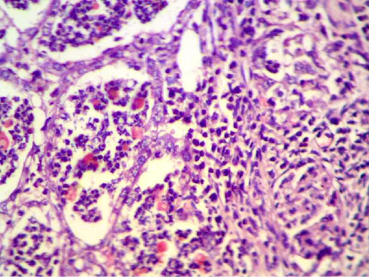
Mammary gland section shows abscess formation inside the mammary alveoli
(H&E 400 X)
Lung section shows sever inflammatory reaction around the small bronchial (bronchiolitis) congestion and destruction of the alveoli. (H&E 200 X)
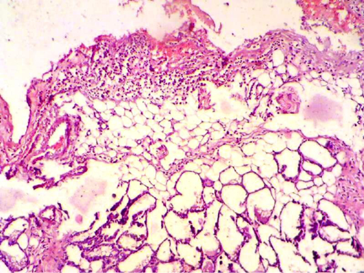
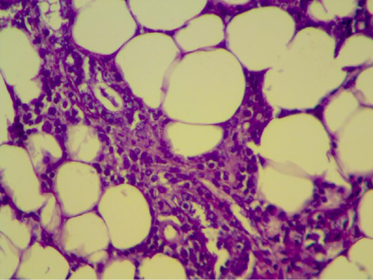
Mammary gland section shows sever inflammatory cell infiltration, haemorrhagic exudate (oedema) and destruction of adipose tissue and mammary alveoli (H&E 200 X)
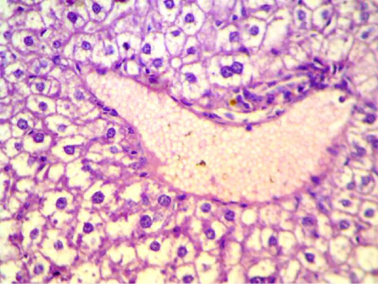
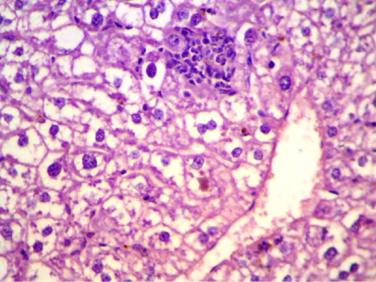
Liver section shows depletion of glycoprotein granules with degeneration and necrosis and inflammatory cell infiltration (H&E 400 X)
Liver section shows depletion of glycoprotein granules with degeneration and necrosis and inflammatory cell infiltration (H&E 200 X)
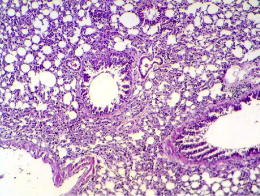
Lung section shows damage of alveoli septa and small bronchial epithelial (H&E 200 X)
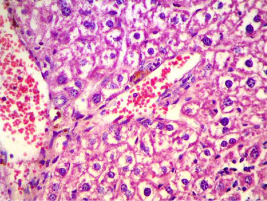
Liver section shows congestion with degeneration and depletion of glycoprotein granules (H&E 400 X)
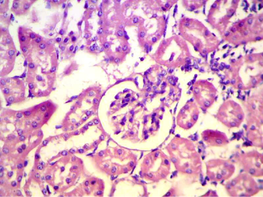
Kidney section shows degeneration, necrosis in renal tubules and inflammatory cell infiltration. (H&E 400 X)
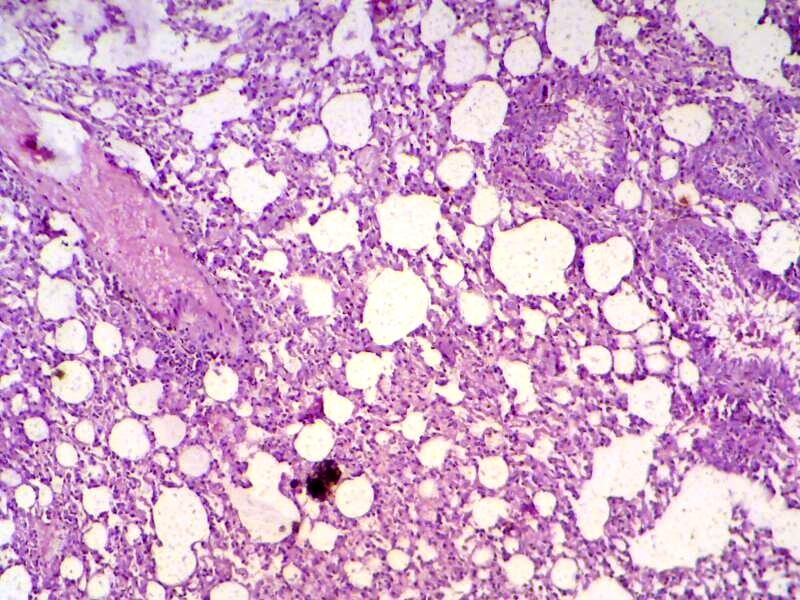
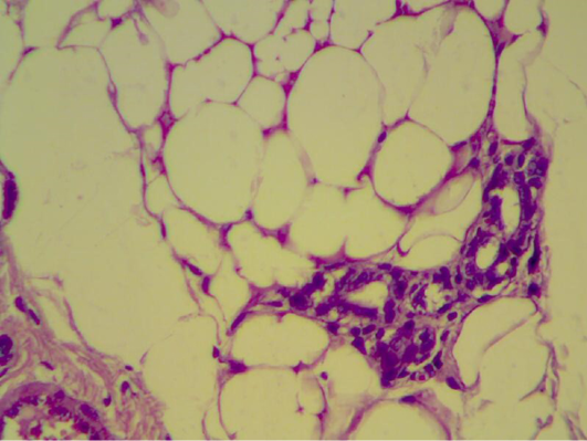
Mammary gland section shows inflammatory reaction around mammary alveoli with destruction of adipose tissue. (H&E 200 X)
Mammary gland section shows look – like normal appearance of mammary tissue. (H&E 400 X)
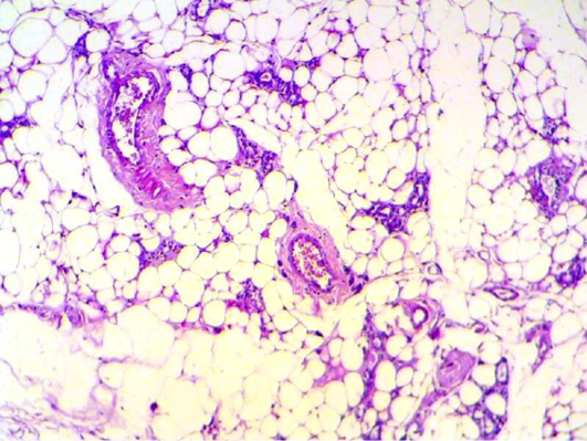
Mammary gland section shows mild inflammatory cell with normal looking adipose tissue. (H&E 200 X) (H&E 400 X)


