Journal of Animal Health and Production
Research Article

Ultrasonography as a Decision Making Tool for Respiratory Affected Calves
Shaimaa Mohamed Gouda
Department of Animal Medicine, Faculty of Veterinary Medicine, Zagazig University, Egypt.
Abstract | The purpose of this study was to clarify the importance of the ultrasonography in assessing the severity of respiratory troubles in calves. For this purpose, 30 calves were investigated in this study. By using scoring system for calves’ respiratory disease, these calves were classified into 2 groups. The 1st group included 15 calves with total scoring less than 4 (normal group) while the 2nd group contained 15 calves with total scoring equal or more than 4 (respiratory affected group). Auscultation and ultrasonographic examination of lung field were applied on both groups. Calves in 1st group were healthy and had no abnormalities in lung either by auscultation or by ultrasound. In the 2nd group, abnormal lung sounds were audible in 11 calves while lung lesions were detected by ultrasound in 7 calves. Furthermore, among 7 calves with lung lesions, 5 had focal lung lesions and 2 had diffuse lung lesions. By receiving the treatment for 5 successive days, 13 calves were responding and the 2 calves with diffuse lung lesion were not responding and euthanized. Necropsy findings of the euthanized calves confirmed the ante-mortem ultrasonographic examination. In conclusion, scoring system for calves’ respiratory disease and chest auscultation are useful tool for detecting the respiratory affected calves in the herd but ultrasonography consider useful tool in evaluation the involvement of lung and the degree of affection.
Keywords | Calves, Respiratory, Ultrasonography, Scoring system, Pneumonia
Editor | Asghar Ali Kamboh, Sindh Agriculture University, Tandojam, Pakistan.
Received | May 12, 2015; Revised | May 24, 2015; Accepted | May 24, 2015; Published | May 30, 2015
*Correspondence | Shaimaa Mohamed Gouda, Zagazig University, Egypt; Email: shaimaagouda81@gmail.com
Citation | Gouda SM (2015). Ultrasonography as a decision making tool for respiratory affected calves. J. Anim. Health Prod. 3(2): 43-47.
DOI | http://dx.doi.org/10.14737/journal.jahp/2015/3.2.43.47
ISSN | 2308–2801
Copyright © 2015 Gouda. This is an open access article distributed under the Creative Commons Attribution License, which permits unrestricted use, distribution, and reproduction in any medium, provided the original work is properly cited.
INTRODUCTION
Respiratory disease is a major economic burden to the calves industry (Bleul, 2009; Pardon et al., 2011). Bovine respiratory disease (BRD) has a multifactorial etiology and develops as a result of complex interactions between environmental factors, host factors, and pathogens (Radostits et al., 2007).
Numerous diagnostic methods are used in the evaluation of different respiratory diseases in bovine, including auscultation, percussion, blood work, radiography, ultrasonography, and more invasive procedures such as aspirations and biopsies (Wilson and Lofstedt, 2008).
A new respiratory scoring system for diagnosis of respiratory diseases considered easy tool for veterinarians and farm personnel (McGuirk, 2008; Poulsen and McGuirk, 2009). Scoring systems are used in clinical and research settings to classify calves as either healthy or sick for the purposes of treatment or analysis.
Auscultation is also considered as a good diagnostic tool especially in calves less than 6 months, because of a narrower chest wall and lung sounds are easily auscultable (Peek, 2005). Unfortunately, it is difficult to assess the extent of damage to lung tissue in bovine bronchopneumonia by clinical investigation alone, thus further tools are required to assess the degree of lung damage (Rabeling et al., 1998).
In veterinary medicine, radiography is also superior to ultrasonography in the identification of diffuse lung diseases (Braun et al, 1997). Ultrasonography of the lungs is established to be the most often applied in diagnosis of lung diseases (Floeck, 2004; Sleeter and Step, 2007; Babkine and Blond, 2009; Scott, 2012). Inflammation and infection of lung resulting in evacuation the air and losing the strong reflector character of normal lung. The reverberation artefact of normal lung changed to a homogenous hypo echoic structure similar to that of liver making it possible to reach an ultrasonographic diagnosis of lung lesions regardless of the clinical state of the animal (Reinhold et al., 2002). The extent of the pneumonia can be evaluated by visualizing either small, localized lesions or lesions that involve the entire part of a pulmonary lobe (Peek, 2005; Scott, 2009). Ultrasonography using a 3.5 MHz sector transducer was not particularly effective as a prognostic/diagnostic tool for early detection of bovine respiratory diseases, but may be useful in targeted populations of animals with respiratory disease of longer duration (Abutarbush et al., 2012).
The current study was planned to use the ultrasonography in evaluation of respiratory affected calves after preliminary evaluation by respiratory scoring system for reaching to a suitable decision.
MATERIALS AND METHODS
Animal Selection
Thirty suckling calves weighed 50- 80 kg, were selected from a herd of calves in Elsalhia farm at Sharkia Governorate- Egypt in December, 2014. After application of scoring system for calves’ respiratory disease, calves were grouped into 2 groups. The 1st group consisted 15 calves with scoring less than 4 and the 2nd group consisted 15 calves had scoring more than 4.
Scoring System for Calves’ Respiratory Disease
This system was described by (Lago et al., 2006; McGuirk, 2008), it depends up on scoring the rectal temperature, cough, nasal discharge, eye and ear scores (Table 1).
Table 1: Calf health scoring criteria (McGuirk, 2008)
|
Calf Health Scoring Criteria |
|||
|
0 |
1 |
2 |
3 |
|
Rectal Temperature |
|||
|
100-100.9 F (37.8-38.2oC) |
101-101.9 F (38.3- 38.8oC) |
102-102.9 F (38.9- 39.3oC) |
≥ 103 F (≥ 39.4oC) |
|
Cough |
|||
|
None |
Induce single cough |
Induced repeated coughs or occasional spontaneous cough |
Repeated spontaneous cough |
|
Nasal discharge |
|||
|
Normal serious discharge |
Small amount of unilateral cloudy discharge |
Bilateral, cloudy or excessive mucus discharge |
Copious bilateral mucopurulent discharge |
|
Eye score |
|||
|
Normal |
Small amount of ocular discharge |
Moderate amount of bilateral discharge |
Heavy ocular discharge |
|
Ear score |
|||
|
Normal |
Ear flick or head shake |
Slight unilateral drop |
Head tilt or bilateral drop |
Auscultation Technique
Auscultation of the lung field was applied on both sides of the chest with Littmann stethoscope (Classic II SE- USA). All area of lung was auscultated in a zigzag manner as described previously by Rosenberger (1990).
Ultrasonographic Examination
The area between the fifth and twelfth intercostal spaces is shaved on the right and left sides in the shape of a triangle that spans the caudal edge of the thoracic limb, the transverse vertebral processes, and a line extending from the elbow to the dorsal corner of the thirteenth rib and following the diaphragm. Each intercostal space was examined after the method described previously by Rabeling et al. (1998). Application of acoustic gel then a linear 6-8 MHz probe with penetration depth 5-10 cm is placed parallel to the ribs (SonoScape- China).
Therapeutic Plan
All calves with scoring equal to or more than 4 were received treatment. The line of treatment included antibiotic, non-steroidal anti-inflammatory (NSAIDs) and multi-vitamins as stated by Radostits et al. (2007). The antibiotic was Tulathromycin (Draxxin®- Pfizer Company) by a single dose of 1ml per 50 kg body weight administered subcutaneously. Flunixin meglumine was the NSAIDs of choice in this investigation (Finadyn solution®- Schering Plough Animal Health Company) by a dose of 2 ml per 45 kg body weight administered intravenously for 5 consecutive days. Finally, the multi-vitamin was ADEVIT-C (ADWIA Company) by a dose of 2 ml per calf administered intramuscular for 5 days.
RESULTS
As shown in Table 2, calves were classified basing on respiratory scoring system into 2 groups. First group included 15 calves with total respiratory scoring less than 4 (Figure 1A), these calves were described as normal .While the second one included 15 calves with total respiratory scoring more than 4 (Figure 1B) and described as respiratory affected calves.

Figure 1: (A) Calf with total respiratory scoring less than 4 and grouped as normal. (B) Calf with total respiratory scoring more than 4 and grouped as respiratory affected.
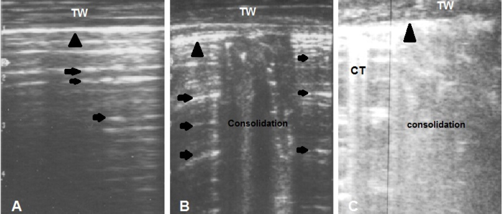
Figure 2: (A) Ultrasonogram of normal lung shows presence of reverberation artifact (arrows) medial to the hyperechoic pleura (arrow head). (B) Focal area of consolidation in between normal lung tissue (mild cases). (C) Diffuse area of consolidation and comet tail (CT) artifact reflect presence of severe disease. TW: Thoracic wall
Auscultation of the lung field of affected calves’ revealed normal vesicular sound of the lung in 4 calves while abnormal lung sounds included wheezes, crackling and exaggerated vesicular sound were audible in 11 cases. The abnormal sound was audible at local area either left or right in 5 calves while was audible all over lung at 6 calves.
Table 2: Total respiratory scoring in examined calves
|
Case number |
Age |
Temperature |
Nasal discharge |
Cough |
Eye or ear |
Total* score |
|
1 |
1 wk |
1 |
0 |
0 |
1 |
2 |
|
2 |
2 wk |
1 |
0 |
0 |
0 |
1 |
|
3 |
3 wk |
2 |
0 |
0 |
0 |
2 |
|
4 |
2 wk |
1 |
0 |
0 |
0 |
1 |
|
5 |
4 wk |
2 |
0 |
0 |
0 |
2 |
|
6 |
3 wk |
0 |
0 |
0 |
0 |
0 |
|
7 |
3 wk |
1 |
1 |
0 |
0 |
2 |
|
8 |
1 wk |
1 |
0 |
0 |
0 |
1 |
|
9 |
2 wk |
1 |
0 |
0 |
0 |
1 |
|
10 |
4 wk |
2 |
0 |
0 |
0 |
2 |
|
11 |
2 wk |
0 |
1 |
0 |
0 |
1 |
|
12 |
3 wk |
2 |
1 |
0 |
0 |
3 |
|
13 |
4 wk |
1 |
1 |
0 |
0 |
2 |
|
14 |
4 wk |
0 |
0 |
0 |
0 |
0 |
|
15 |
3 wk |
1 |
0 |
0 |
0 |
1 |
|
16 |
3 wk |
3 |
2 |
2 |
1 |
8 |
|
17 |
1 wk |
3 |
2 |
2 |
0 |
7 |
|
18 |
3 wk |
3 |
1 |
3 |
0 |
7 |
|
19 |
1 wk |
3 |
2 |
1 |
2 |
8 |
|
20 |
1 wk |
3 |
2 |
3 |
0 |
8 |
|
21 |
2 wk |
3 |
1 |
2 |
0 |
6 |
|
22 |
2 wk |
3 |
1 |
0 |
0 |
4 |
|
23 |
4 wk |
3 |
1 |
1 |
0 |
5 |
|
24 |
3 wk |
2 |
2 |
1 |
0 |
5 |
|
25 |
2 wk |
3 |
2 |
3 |
0 |
8 |
|
26 |
3 wk |
3 |
3 |
1 |
0 |
7 |
|
27 |
3 wk |
3 |
3 |
1 |
0 |
7 |
|
28 |
4 wk |
3 |
1 |
1 |
0 |
5 |
|
29 |
3 wk |
3 |
2 |
1 |
0 |
6 |
|
30 |
4 wk |
3 |
3 |
2 |
0 |
8 |
* Total respiratory scoring from case 1 to case 15 are less than 4 and grouped as normal while from case 16 to case 30 are more than 4 and grouped as respiratory affected calves
Ultrasonography revealed presence of normal character of healthy lungs in 23 calves (15 of 1st group and 8 in 2nd group) and presence of consolidated area in 7 calves. The healthy lung appeared with double echogenic pleural lines and reverberations artefact represent the normal lung (Figure 2A). The affected lung areas appeared with hypoechoic part and comet tail artefact that replaced the reverberation artifact of normal lung. This area was localized only in small part in 5 cases (Figure 2B) and diffuse included the most part of lung in 2 cases (Figure 2C).
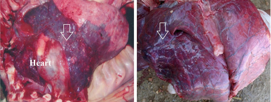
Thirteen calves improved clinically after the therapeutic trial, and become with total respiratory score less than 4 while the 2 calves with diffuse lung lesion did not respond and were euthanized (Figure 3).
DISCUSSION
Respiratory diseases are the principal source of economic loss among calves’ herds all over the world. Many diagnostic tools were used in evaluation of such diseases. In the present study, 3 tools were used in evaluation of 30 calves in a farm with an outbreak from respiratory diseases.
As an initial evaluation by application of respiratory scoring system, calves were grouped into healthy and respiratory affected calves but not suitable for evaluation the severity of the disease. This result was in accordance to Poulsen and McGuirk (2009).
Abnormal lung sounds by auscultation were audible on lung field at 11 cases. Comparatively, lung lesions were observed by ultrasonography in 7 cases. It means that 4 calves had abnormal lung sounds with no lung lesion by ultrasonography. This may attribute to that the lung of these calves is not involved and the abnormal lung sounds are due to bronchitis or due to involvement of the lung but at early stage. This result is in agreement to Abutarbush et al. (2012) who stated that ultrasonography is useful only in late stage of lung involvement and not useful in early prediction of lung disease. This result indicates also that when the lung is involved by ultrasonography, the disease is more severe. Therefore, ultrasonography consider as a prognostic tool. Similar result has been reported previously by Scott (1998) and Floeck (2004) in adult cattle.
Regarding the ultrasonography of normal lung, it appeared with hyperechoic linear image, often with equally-spaced reverberation artifacts below this line as previously described by Reinhold et al. (2002). The reverberations results from reflection of ultrasound waves by air-filled lung tissue, and forth on the border between tissue and lung parenchyma, simulating hyperechoic pulmonary structures in the form of reverberation artifacts (Blond and Buczinski, 2009). On the other hand, the affected lung appeared with hepatisation or consolidation at which the reverberations are replaced by hypoechoic structure, resulting in an ultrasonographic appearance similar to that of a liver (Rabeling et al., 1998). Moreover, comet-tail artifacts appeared as bright, ray-like stripes distal to a small hyperechoic structure whose impedance difference to the surrounding tissue is very large. They correspond to intensive multiple reflections (Braun et al., 1997). In accordance with the present study, this pattern was designated as a severe lesion when it covered an extensive or diffuse area of lung tissue (no=2) and mild to moderate abnormalities when it covered small or focal area of the lung (n=5). This result is in consistent with those obtained previously (Klein, 1999).
The result of therapeutic trials revealed a clinical response in mild cases of lung involvement (n=5) and respiratory affected calves without lung involvement (no=8) while the 2 cases with diffuse lung lesion not respond to the treatment.
CONCLUSION
Ultrasonography consider as a decision making tool for respiratory affected calves, because it made it possible to determine the location and extent of the lung lesions accurately.
CONFLICT OF INTEREST
Author declares no conflict of interest.
Aknowledgement
I would like to thank all Veterinary doctors in Elsalhia Farm, Sharkia, Egypt for their support during conduction of this work.
REFERENCES


