Journal of Animal Health and Production
Research Article
J. Anim. Health Prod. 3 (2): 43 -47

Figure 1
(A) Calf with total respiratory scoring less than 4 and grouped as normal
(B) Calf with total respiratory scoring more than 4 and grouped as respiratory affected
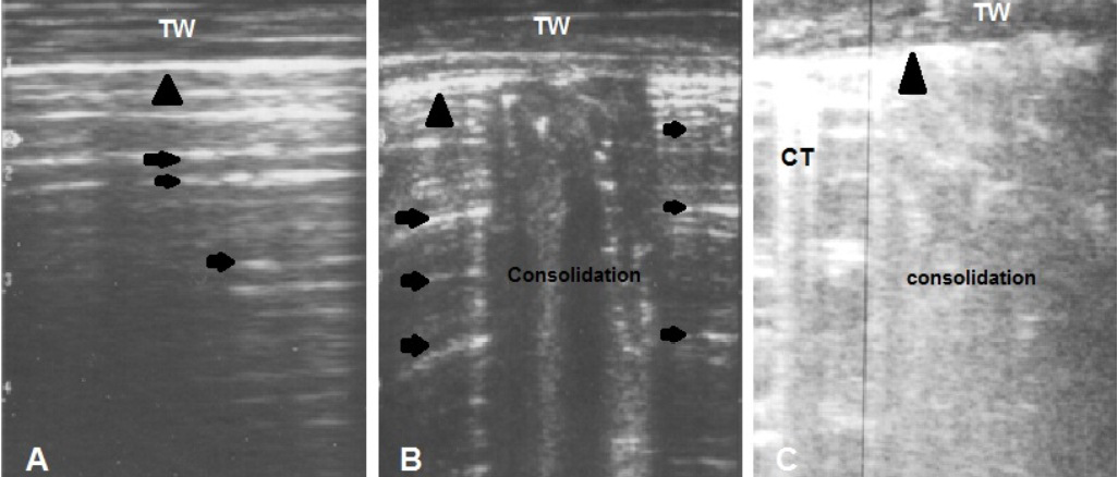
Figure 2
(A) Ultrasonogram of normal lung shows presence of reverberation artifact (arrows) medial to the hyperechoic pleura (arrow head)
(B) Focal area of consolidation in between normal lung tissue (mild cases)
(C) Diffuse area of consolidation and comet tail (CT) artifact reflect presence of severe disease. TW: Thoracic wall
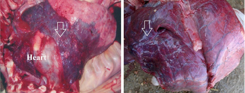
Figure 3
Necropsy finding of two calves with diffuse lung consolidation (arrow)


