Journal of Animal Health and Production
Case Report
Journal of Animal Health and Production 2 (2): 28 – 30Bilateral Mastectomy for Successful Management of Chronic Suppurative Mastitis in a Black Bengal Doe (Capra Hircus)
Shongsir Warson Monsang1*, Saumen Kanti Pal2, Mritunjay Kumar1, Joyabrata Roy1, Charan Singh Sharma3, Mangsatabam Norjit Singh4,
- Department of Teaching Veterinary Clinical Complex, College of Veterinary Sciences & A.H., R. K. Nagar, Tripura (W), India
- Department of Pharmacology and Toxicology, College of Veterinary Sciences & A.H., R. K. Nagar, Tripura (W), India
- Department of Veterinary Medicine, College of Veterinary Sciences & A.H., R. K. Nagar, Tripura (W), India
- Department of Parasitology, College of Veterinary Sciences & A.H., R. K. Nagar, Tripura (W), India
*Corresponding author: warsonmonsang@gmail.com
ARTICLE CITATION:
Monsang SW, Pal SK, Kumar M, Roy J, Sharma CS, Norjit Singh MN (2014). Bilateral mastectomy for successful management of chronic suppurative mastitis in a black bengal doe (Capra hircus). J Anim Health Prod. 2 (2): 28 – 30.
Received: 2014–02–24, Revised: 2014–05–10, Accepted: 2014–05–17
The electronic version of this article is the complete one and can be found online at
(
http://www.nexusacademicpublishers.com/table_contents_detail/11/324/html
)
which permits unrestricted use, distribution, and reproduction in any medium, provided the original work is properly cited
ABSTRACT
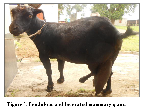
A case of chronic suppurative mastitis in an eight years old Black Bengal doe suffering for the past eight or nine months was presented with the history of non responsiveness to any medicinal treatments subjected to worsen the condition to incurable grade. On examination, the mammary gland was pendulous, lacerated, mildly fibrotic and non–painful which hampers animal walk. The inguinal lymph nodes were also swollen on careful hand palpation. Haematology revealed lower level of haemoglobin (7g/dL) and normal total leukocyte count (9400/cmm) along with relative neutrophilia (78%). Based on the non–responsive and chronic nature of the condition, radical surgery was planned for total ablation of the mammary gland. Surgical anaesthesia was achieved with the combination of diazepam and ketamine in 1:2 ratios without any complications. Complete healing of the wound was observed on the 2nd week.
Mastitis reflects the inflammation of the mammary gland, which may occur due to any bacterial infection secondary to teat injury or poor management (Marogna et al., 2012). It is more frequent in dairy and meat goats raised under intensive and semi–intensive management practices. Mastitis is generally associated with poor hygienic practices and caused by the bruising of mammary tissue or teats from traumas, nursing, fly bites, or other wounds to the skin that provide an important barrier to infection. It may also be associated with viral, bacterial or fungi and their toxins.
Chronic mastitis develops from an untreated case of acute mastitis which is manifested as formation of abscess within the mammary parenchyma (Scott, 2007). Early recognition and prompt treatment are important for limiting tissue damage and production losses. Practically, most of the mastitis cases are well treated with antibiotic injections as well as antibiotic teat infusions. In cases where medicinal treatment is obsolete, unilateral or bilateral mastectomy is recommended as a pain relieving procedure for extensive lesions involving udder and in cases of chronic mastitis, gangrenous lesions or neoplasia (Cable et al., 2004). The present case report describes bilateral mastectomy for the management of chronic suppurative mastitis in a suffering mastitic Black Bengal doe.
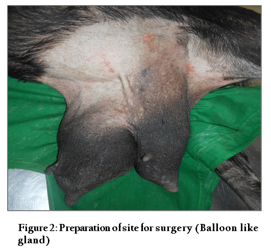
An eight years old matured Black Bengal doe was presented to the Teaching Veterinary Clinical Complex, R.K.Nagar, Tripura, with the history of chronic mastitis. Anamnesis revealed recurrent mastitis non responsive to any medicinal treatment persisting for the past eight or nine months. On clinical examination, the mammary gland was pendulous and lacerated reaching the floor of the ground which hampers the animals walk (Figure 1). The udder was filled with watery fluid like milk having the appearance of fluid–filled balloon (Figure 2) and laceration was also observed. The inguinal lymph nodes were also found to be enlarged.
All the clinical parameters were within the normal physiological limits and the animal was having normal appetite. Mild fibrotic consistency of mammary gland was felt without yielding any pain on palpation. Haematology revealed lower level of haemoglobin (7g/dL) and normal total leukocyte count (9400/cmm) along with relative neutrophilia (78%). Based on the non–responsive and chronic nature of the condition, radical surgery was planned for total ablation of the mammary gland.
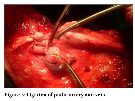
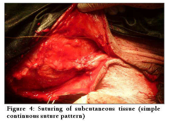
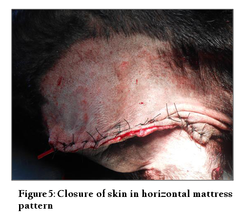
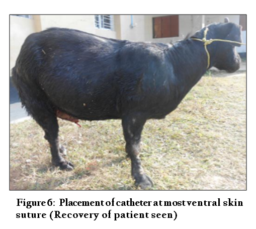
Preparation of the animal consisted of fasting and withholding of feed and water for 12 hours prior to surgery. Animal was sedated with Siquil (Triflupromazine HCl) @0.2 mg/kgbw, IV (Balagopalan, 1990) and controlled in lateral recumbency after general anaesthesia with ketamine (4mg/kgbw, IV) (Hall and Clarke, 1991). Maintenance was done with a mixture of diazepam and ketamine in 1:2 ratios mixed in a single syringe infused intravenously (Hall and Clarke, 1991). Under aseptic environment, an incision was given at the base of the gland and a blunt dissection was made to undermine the skin to separate the tissues properly. The external pudic artery and vein, perineal artery and large subcutaneous vein were isolated and ligated to avoid haemorrhage (Figure 3). During surgery, mild bleeding from subcutaneous bleeders was controlled by ligation with chromic catgut (1–0). After total removal of the mammary gland, the subcutaneous tissue were sutured with chromic catgut 2–0 in simple continuous suture pattern (Figure 4) and the skin was sutured with black braided silk in horizontal mattress pattern (Figure 5). Postoperative care included Cefotaxime (1g, IM, BID) and Flunimeg (1.5mL, IM, OD) for 5 and 3 days, respectively. Antiseptic dressing with povidone iodine and Charmil ointment was advised regularly till healing is completed. Skin sutured removal was advised after 10 days of post surgery. Additionally, a fenestrated cathether was fixed at the most ventral skin sutured to drain out any fluid or seroma (Figure 6). The fluid sample collected before the surgery was cultured for bacterial and sensitivity tests and the results were found to be negative for any bacterial species. After 15 days, the animal recovered fully with complete healing of surgical wound and the skin sutures were also removed.
In a herd system, chronically infected mastitic doe’s are recommended for culled as they serve as a source of infection to other healthy herd. In cases where the infection is systemic, suitable local or parenteral antibiotics along with antihistamines and antiinflammatory are projected to remove the cause. If mastitis in localized to the udder, treatment by first "milking out" the affected gland is best considered.
Radical mastectomy (unilateral or bilateral) is a salvage procedure and indicated in cases of chronic suppurative mastitis, gangrenous mastitis and neoplastic or hyperplastic conditions of the udder (Andreasen et al., 1993; El–Maghraby, 2001; Cable et al., 2004). Chronic mastitis associated with single or multiple abscessation has been reported in sheep (Scott, 2007). Development and growth of such abscesses with proliferation of surrounding fibrous tissue leads to gross enlargement of the udder which is very painful and presents a welfare concern to the animal. In the present case, the doe had a chronic swelling with abscesses within the mammary parenchyma along with mild fibrosis. Moderate anaemia could be due to frequent hemorrhages from the lacerations and relative neutrophilia could be associated with suppurative condition of udder.
Taking into considerations the chronic and non–responsive nature of mastitis, decision to amputate the whole mammary gland was undertaken to relieve the discomfort of the animal while walking and also to improve the health of the suffering doe so that it could be beneficial for the owner to generate income after being sold for meat. In conclusion, bilateral mastectomy is a safe and alternate procedure to treat untreatable conditions of the udder especially in cases of chronic suppurative mastitis in small ruminants.
REFERENCES
Andreasen CB, Huber MJ, Mattoon JS (1993). Unilateral fibroepithelial hyperplasia of the mammary gland in a goat. J. Am. Vet. Med Ass. 202: 1279–1280.
PMid:8496086
Balagopalan TP, Nayar KNM, George PO (1990). Effect of guaiacolate ether alone and in combination with Triflupromazine hydrochloride and thiopentone sodium in goats. Indian Vet. J. 67(8): 739–742.
Cable CS, Peery K, Fubini SL (2004). Radical mastectomy in 20 ruminants. Vet. Surg. 33: 263–266.
http://dx.doi.org/10.1111/j.1532-950X.2004.04038.x
PMid:15104633
El–Maghraby HM (2001). Comparison of two surgical techniques for mastectomy of goats. Small Rum. Res. 40: 215–221.
http://dx.doi.org/10.1016/S0921-4488(01)00173-0
Hall LW, Clarke KW (1991). Anesthesia of the sheep, goat and other herbivores. 9th edn., ELBS Bailliere Tindall, London pp. 260–274.
Marogna G, Rolesu S, Lollai S, Tola S, Leori G (2010). Clinical findings in sheep farms affected by recurrent bacterial mastitis. Small Rum. Res. 88 (2–3), 119–125.
http://dx.doi.org/10.1016/j.smallrumres.2009.12.019
Scott PR (2007). Sheep Medicine. 1st edn., Manson Publishing, The Veterinary Press, London pp. 274–276.
PMid:17630111


