Journal of Animal Health and Production
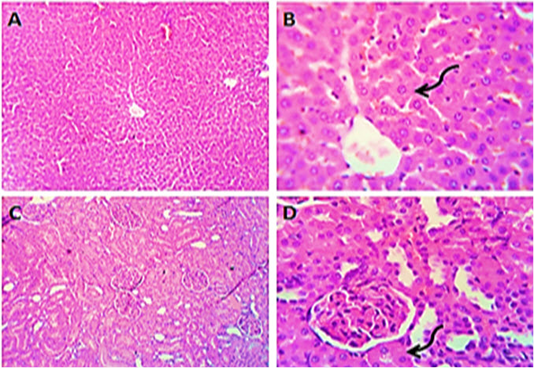
Photomicrographs of liver (A&B) and kidney (C&D) sections from control group (group1) showing normal histo-morphologic structures with mild degenerative changes in some hepatocytes and some of the renal tubular epithelium (curved arrows). H&E. Magnification x100 (A, C) & x400 (B, D).
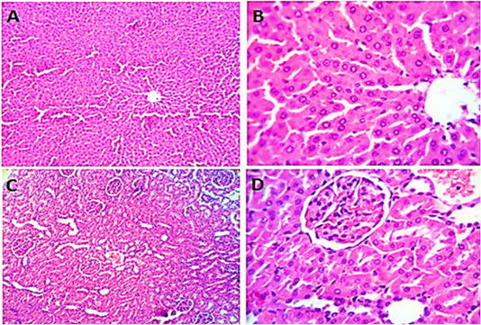
Photomicrographs of liver sections (group2) from male rats given olive oil showing normal hepatic parenchyma with preserved lobular pattern, portal triads structures, vascular tree, kupffur cells and stromal component (A, B). Kidney sections (group2) of male rats received olive oil showing normal nephron units with preserved glomerular and tubular structures, normal limits with normal histomorphology of the blood vessels and stroma (C, D). H&E. Magnification x400.
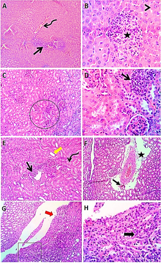
Photomicrographs of liver sections (group3) from male rats received levofloxacin showing moderate congestion of portal blood vessels (curved arrow) with round cells aggregations (open arrow), focal interstitial round cells aggregation (star) with degenerative changes (arrow head) in some hepatocytes (A & B). Kidney sections (group3) of male rats exposed to levofloxacin showing lymphocytic interstitial aggregations (open arrows), perivascular edema (star) sometimes with minute hemorrhages (closed arrow), dilated renal tubules (thick yellow arrow) in both cortex and medulla with intratubular hyaline cast (curved arrow). The renal pelvis and the renal papillae showing lymphocytic pyleitis (square) with superficial ulceration of the lining epithelium (red thick arrow) and moderate subepithelial lymphocytic aggregation (black thick arrow) (C- H). H&E. Magnification x100 (A, C, E, G) & x400 (B, D, F, H).
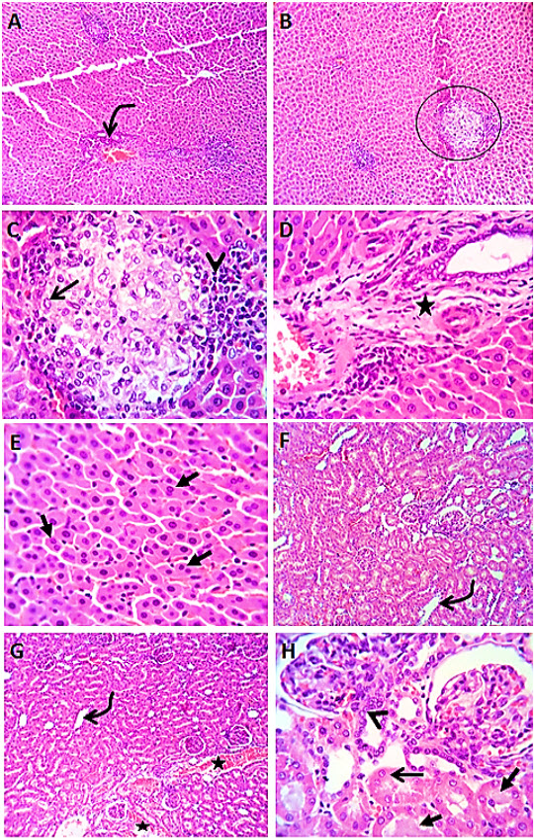
Photomicrographs of liver sections (group4) from male rats given vitamin E before the administration of levofloxacin by 2hours showing lymphocytic aggregation in the portal areas (curved arrow) with healing reaction in both the portal area (star) and the interstitial tissue (circle), with replacement of the lymphocytes (arrow head) by macrophages (open arrow) or fibroblasts. Most of the hepatic parenchyma showing active hepatocytes with increased number of binucleated cells (closed arrows) (A-E). Kidney sections (group4) showing mild congestion (stars) of renal blood vessels, dilatation of some cortical and medullary tubules (curved arrows) beside regenerative attempts in some tubules (arrow head). A few tubules showing cloudy swelling (open arrow) and /or hyaline casts (closed arrows) (F- H). H&E. Magnification x100 (A, B) & x400 (C, D, E, F, G, H).
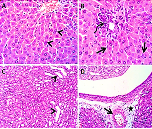
Photomicroraph of liver sections (group5) from male rats given Panax ginseng before the administration of levofloxacin by 2hours showing some binucleated hepatocytes (arrow heads) with activated kupffer cells (open arrow), beside mild lymphocytic reaction (curved arrow) in the portal area (A &B). Kidney sections (group5) showing mildly congested renal blood vessels (open arrow) with perivascular edema (star) and some renal tubules in both cortex and medulla are dilated (arrow heads) (C & D). H&E. Magnification x100 (C, D) & x400 (A, B).


